


Neurotrophic Keratitis: When the Cornea Loses Sensation

Neurotrophic keratitis develops when the nerves supplying your cornea become damaged, reducing sensation and making corneal problems harder to notice.
Your condition can worsen without much pain and may lead to persistent surface damage or corneal thinning. Early diagnosis and specialist monitoring can help protect your cornea.
What Is Neurotrophic Keratitis?
Neurotrophic keratitis can develop when the sensory nerves supplying your cornea are damaged, reducing its sensitivity. These nerves also help control your blinking, tear production and corneal healing.
Your cornea may therefore become damaged without causing the usual pain, watering or irritation. This can allow neurotrophic keratitis to progress quietly, so early assessment is important.
Why Does the Cornea Need Sensation?
Your corneal nerves help protect your eye by triggering blinking and tear production when your cornea is irritated. They also support the normal maintenance and healing of your corneal surface.
Your reduced corneal sensation can therefore make eye damage harder to notice. You may have little pain or watering even while your cornea is becoming damaged.
Evidence Note
Research shows that corneal sensory nerves do more than allow you to feel pain or irritation. They also contribute to blinking, tear-film stability and the maintenance and healing of the corneal epithelium. When trigeminal nerve function is reduced, these protective mechanisms can become impaired, making the cornea more vulnerable to epithelial breakdown and delayed healing.
This helps explain why neurotrophic keratitis can progress with surprisingly little discomfort. Reduced sensation may remove the normal warning signal that would otherwise prompt you to seek treatment, so an apparently comfortable eye can still have significant corneal surface damage. Early assessment of corneal sensation and the ocular surface is therefore important when neurotrophic keratitis is suspected.
What Causes Neurotrophic Keratitis?
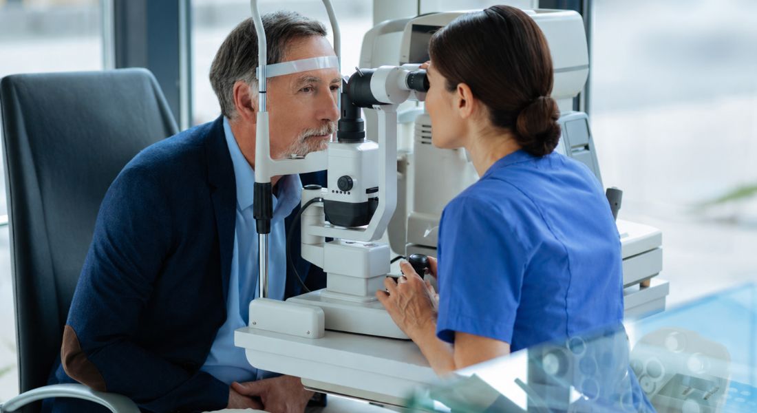
Neurotrophic keratitis can develop when the nerves supplying your cornea are damaged. Herpes infections, diabetes, previous eye surgery, trauma and some neurological conditions can all contribute.
Your corneal nerves can also be affected by burns, certain nerve procedures or long-term use of some eye medicines. Finding the underlying cause can help your ophthalmologist protect your cornea and guide your treatment.
Can Herpes Simplex or Shingles Cause Loss of Corneal Sensation?
A previous herpes simplex or shingles infection affecting your eye can damage the nerves supplying your cornea. Your corneal sensation may remain reduced even after the infection or rash has settled.
- Nerve damage: Herpes infections can affect the corneal nerves and reduce your ability to feel the surface of your eye.
- Reduced pain sensation: New corneal damage may cause little or no pain when corneal sensation is reduced.
- Symptoms may be subtle: You may notice blurred vision, dryness or other surface changes rather than significant discomfort.
- Previous infections matter: Your ophthalmologist may ask about previous herpes infections when investigating reduced corneal sensation.
Reduced corneal sensation can increase the risk of unnoticed corneal damage. Regular eye assessment can help your ophthalmologist monitor the cornea and identify problems early.
Can Diabetes Cause Neurotrophic Keratitis?
Your diabetes can affect the nerves in your cornea and reduce its sensation. This does not mean you will develop neurotrophic keratitis, but reduced sensation can make corneal problems harder to notice.
Your ophthalmologist may check your corneal sensation if you have diabetes and a persistent surface problem with little discomfort. Managing your diabetes well is also important for your overall nerve health.
Can Previous Eye Surgery Cause Neurotrophic Keratitis?
Your previous eye surgery can sometimes affect the nerves in your cornea and reduce its sensation. Procedures such as LASIK and PRK can temporarily reduce corneal sensation, although sensation usually improves over time.
Your risk depends on the type of surgery and how your cornea heals. If you develop ongoing dryness, surface damage or surprisingly mild symptoms after surgery, your ophthalmologist may check your corneal sensation.
What Are the Symptoms of Neurotrophic Keratitis?
Neurotrophic keratitis may cause blurred vision, mild redness, watering or dryness, but you may have little or no pain because your cornea has reduced sensation.
Your eye can therefore look or feel relatively comfortable even when the corneal surface is damaged. If you develop new blurred vision or redness, you should have your eye assessed rather than assuming it is harmless.
Why Can Neurotrophic Keratitis Be Difficult to Recognise?
Neurotrophic keratitis can be difficult to recognise because your symptoms may seem mild even when your cornea is significantly damaged. Reduced corneal sensation means you may feel little pain or watering from a surface injury.
Your ophthalmologist may still find corneal damage during an examination. Regular eye checks can be important when your corneal sensation is reduced, especially if you have previously had a corneal ulcer or persistent surface defect.
What Are the Three Stages of Neurotrophic Keratitis?
Neurotrophic keratitis is commonly divided into three stages. In Stage 1, your corneal surface develops mild changes without an open defect. In Stage 2, your cornea develops a persistent epithelial defect that does not heal normally.
In Stage 3, your corneal damage becomes deeper and may cause an ulcer, thinning or, in severe cases, perforation. Early treatment aims to protect your cornea and prevent the condition from progressing to the later stages.
Research Insight
Research traditionally describes neurotrophic keratitis using the three-stage Mackie classification, progressing from early epithelial abnormalities to a persistent epithelial defect and then to corneal ulceration, stromal melting and possible perforation. More recent work has highlighted that neurotrophic keratopathy can involve a wider range of epithelial and stromal changes, making early assessment of corneal sensation particularly important.
The key clinical principle is that progression may occur without the degree of pain expected from the amount of corneal damage. This means treatment should not be based only on symptoms; your ophthalmologist also needs to examine the corneal surface, measure or assess sensation and monitor whether the epithelium is healing.
What Happens in Stage 1 Neurotrophic Keratitis?
Stage 1 neurotrophic keratitis can make your corneal surface look unhealthy even though you do not yet have a persistent open wound. You may have surface staining, an unstable tear film or other early changes while your corneal sensitivity is reduced.
Your treatment aims to protect your cornea before a persistent defect develops. Your ophthalmologist may recommend preservative-free lubricating drops, ointment or treatment for your dry eye or eyelid problems, depending on what you need.
What Happens in Stage 2 Neurotrophic Keratitis?
Stage 2 neurotrophic keratitis means your corneal surface has developed a persistent defect that is not healing normally. You may still feel little pain because your corneal sensation is reduced.
Your treatment aims to help your cornea heal and prevent infection or deeper damage. Your ophthalmologist may recommend intensive lubrication, specialist eye drops or other treatments, with close follow-up to monitor your recovery.
What Happens in Stage 3 Neurotrophic Keratitis?
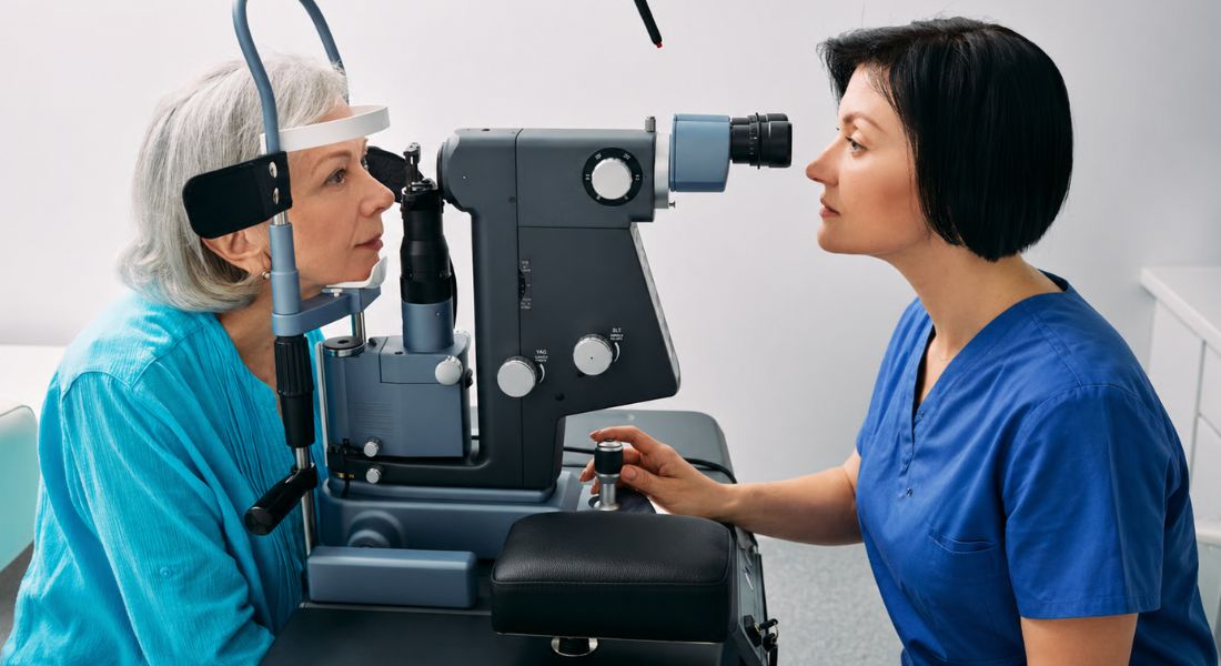
Stage 3 neurotrophic keratitis is the most serious stage because your cornea has developed deeper damage. Your persistent defect can progress to an ulcer, thinning or melting, which can put your eye at risk of perforation.
Your treatment may need to become more intensive to protect the structure of your eye. In severe cases, your ophthalmologist may recommend treatments such as tissue adhesive, an amniotic membrane or corneal surgery.
Stages of Neurotrophic Keratitis
| Stage | What happens to your cornea | Typical features | Main treatment aim |
| Stage 1 | The corneal surface becomes abnormal but there is no persistent open defect | Punctate epithelial changes, reduced tear-film stability and surface irregularity | Protect and stabilise the corneal surface and prevent progression |
| Stage 2 | A persistent epithelial defect develops and does not heal normally | Open epithelial defect with reduced corneal sensation and risk of deeper damage | Promote epithelial healing and prevent infection or further stromal damage |
| Stage 3 | Deeper corneal damage develops | Corneal ulceration, thinning, stromal melting and possible perforation | Protect the structural integrity of the eye and prevent perforation |
How Is Corneal Sensation Tested?
Your ophthalmologist can test your corneal sensation by gently touching your cornea and comparing your response with your other eye. Your doctor may use an esthesiometer for a more precise assessment.
Your sensation should be tested before anaesthetic drops are used because they temporarily numb your eye. Your ophthalmologist will also check your cornea and tear film.
Clinical Tip
If your ophthalmologist is assessing reduced corneal sensation, tell them about any previous herpes infection, shingles affecting your eye, diabetes, corneal surgery, trauma or neurological problems. These details can help identify possible reasons why the nerves supplying your cornea may have been affected.
You should also mention if your eye feels surprisingly comfortable despite redness, blurred vision or a known corneal surface problem. Reduced pain does not necessarily mean that the cornea is healthy, so your examination and sensation testing are important when symptoms appear mild compared with the visible surface changes.
What Other Tests May Be Needed?
Your ophthalmologist may use fluorescein dye and a slit lamp to check your cornea for surface damage, inflammation or scarring. Your tear production, eyelid closure and blinking may also be assessed.
Your doctor may recommend further tests, such as corneal imaging, depending on your findings. Your medical history can also help identify why your corneal sensation has been reduced.
How Is Neurotrophic Keratitis Treated?
Your treatment depends on the stage of your neurotrophic keratitis and what has reduced your corneal sensation. Your ophthalmologist may use preservative-free lubricating drops or ointments and treat problems such as dry eye, blepharitis or incomplete eyelid closure.
Your treatment may become more intensive if your cornea is damaged, including specialist contact lenses, serum tears or other procedures. The main aim is to help your cornea heal and prevent further damage.
What Is Cenegermin and Can It Help?
Treatment with cenegermin may help your cornea heal by supporting the nerves and surface of your eye. It is used for moderate or severe neurotrophic keratitis and is given as frequent eye drops under specialist supervision.
Your ophthalmologist can explain whether cenegermin is suitable for you and whether it is available through your treatment pathway. Your options will depend on the severity of your condition and your individual circumstances.
UK Guidance Note
Cenegermin is a specialist nerve growth factor eye-drop treatment for adults with moderate or severe neurotrophic keratitis. It is given under specialist supervision and may help support corneal healing. Your ophthalmologist can explain whether it is suitable for you and what treatment options are available through your care pathway.
Your treatment options will depend on the severity of your neurotrophic keratitis, the cause of your reduced corneal sensation and the treatment pathway available to you. Your ophthalmologist can explain whether cenegermin or another treatment is appropriate in your circumstances.
Can Neurotrophic Keratitis Require Surgery?
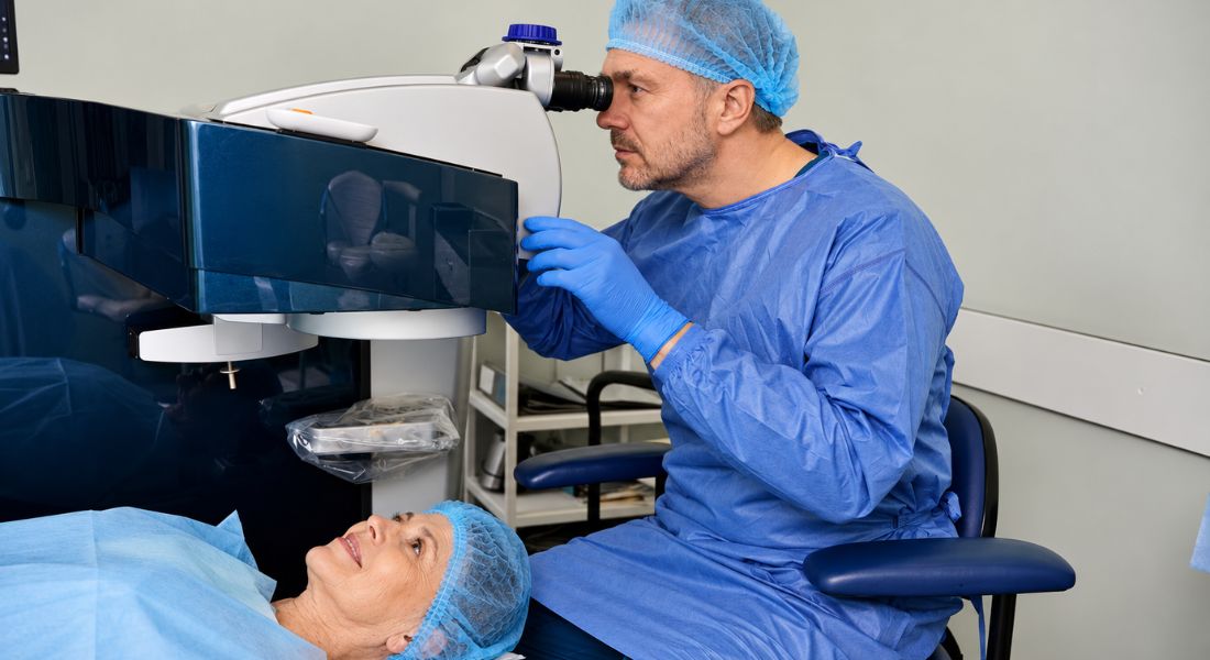
Neurotrophic keratitis may sometimes require surgery when your cornea cannot heal or stay protected with other treatments. Your ophthalmologist may consider procedures such as tarsorrhaphy to help protect your cornea if your eyelid does not close properly.
Severe corneal damage or perforation may require tissue adhesive or a corneal transplant. In selected cases, your specialist may also consider corneal neurotisation to help restore corneal sensation and support corneal healing.
Myth vs Fact
| Myth | Fact |
| Severe corneal damage always causes severe eye pain. | Neurotrophic keratitis can cause significant corneal damage with surprisingly little pain because corneal sensation is reduced. |
| If your eye feels comfortable, your cornea is probably healthy. | A relatively comfortable eye can still have epithelial damage or a persistent defect when corneal sensation is reduced. |
| Neurotrophic keratitis only affects people who have had herpes infections. | Herpes is an important cause, but diabetes, surgery, trauma, neurological conditions and other causes of trigeminal nerve damage can also contribute. |
| Neurotrophic keratitis only becomes serious when an ulcer develops. | Early epithelial changes can progress to persistent defects, ulceration, thinning and perforation if the condition is not recognised and managed. |
| Testing corneal sensation is unnecessary if your vision is good. | Corneal sensation can provide important information because reduced sensation may exist before severe visual loss develops. |
| Treatment is only needed when the eye becomes painful. | Treatment aims to protect the cornea and prevent progression, even when symptoms are mild or pain is absent. |
Key Takeaways
- Neurotrophic keratitis develops when damage to the nerves supplying your cornea reduces corneal sensation.
- Reduced sensation can make corneal damage difficult to notice because pain may be mild or absent.
- Herpes infections, diabetes, previous eye surgery, trauma and neurological conditions can contribute to corneal nerve damage.
- Neurotrophic keratitis is traditionally divided into three stages, progressing from surface changes to persistent epithelial defects and then ulceration or deeper damage.
- Corneal sensation testing is an important part of diagnosis and assessment.
- Your ophthalmologist may also examine the corneal surface, tear film, eyelid closure and healing response.
- Treatment aims to protect the cornea, support epithelial healing and prevent progression.
- Lubrication and other protective treatments may be used, while more advanced disease may require specialist or surgical treatment.
- Cenegermin is a specialist treatment for moderate or severe neurotrophic keratitis, and your ophthalmologist can explain whether it is appropriate and available through your treatment pathway.
- Early diagnosis and regular specialist monitoring can help reduce the risk of persistent defects, corneal thinning and perforation.
Frequently Asked Questions
- What is neurotrophic keratitis?
Neurotrophic keratitis is a condition where the sensory nerves supplying your cornea become damaged, reducing corneal sensation. This can make corneal injury harder to notice and may allow surface damage to progress with little pain. - What causes neurotrophic keratitis?
Neurotrophic keratitis can be caused by damage to the corneal nerves from herpes infections, diabetes, previous eye surgery, trauma or certain neurological conditions. Burns, some nerve procedures and long-term use of certain eye medicines can also contribute. - What are the symptoms of neurotrophic keratitis?
You may have blurred vision, mild redness, watering or dryness, but surprisingly little pain. Reduced corneal sensation can mean that your eye feels relatively comfortable even when the corneal surface is damaged. - Can herpes simplex or shingles cause neurotrophic keratitis?
Yes. A previous herpes simplex or shingles infection affecting your eye can damage the nerves supplying your cornea and leave sensation reduced. This can make later corneal damage more difficult to notice. - What are the three stages of neurotrophic keratitis?
Stage 1 involves early corneal surface changes without a persistent open defect. Stage 2 involves a persistent epithelial defect that does not heal normally, while Stage 3 involves deeper damage such as an ulcer, thinning or, in severe cases, perforation. - How is corneal sensation tested?
Your ophthalmologist can gently test your corneal sensation and compare your response between your eyes. An esthesiometer may also be used to measure sensation more precisely. - How is neurotrophic keratitis treated?
Treatment depends on the stage and cause of your condition. It may include preservative-free lubricating drops or ointments, treatment for dry eye or eyelid problems, specialist contact lenses, serum tears and other treatments to help your cornea heal. - Can cenegermin help treat neurotrophic keratitis?
Yes. Cenegermin is a specialist eye-drop treatment that may help support corneal healing and nerve function in moderate or severe neurotrophic keratitis. Your ophthalmologist will decide whether it is suitable for you. - Can neurotrophic keratitis require surgery?
Yes. Severe or non-healing disease may require procedures such as tarsorrhaphy, tissue adhesive, an amniotic membrane or corneal surgery. In selected cases, corneal neurotisation may also be considered to help restore corneal sensation and support corneal healing. - Why is early treatment important for neurotrophic keratitis?
Your reduced corneal sensation can allow damage to progress without the usual warning of pain. Early diagnosis, specialist monitoring and treatment can help your cornea heal and reduce the risk of ulcers, thinning or perforation.
Final Thoughts: Neurotrophic Keratitis
Neurotrophic keratitis can progress without causing the level of pain you might expect because reduced corneal sensation makes damage harder to notice. Early diagnosis and appropriate treatment can help protect your cornea, support healing and reduce the risk of complications such as persistent defects, ulcers, thinning or perforation.
If you would like a detailed assessment of your eye health, you can contact the Eye Clinic London team to arrange a consultation.
References:
- Eio, E., Yu, M., Liu, C., Lee, I.X.Y., Wong, R.K.T., Wong, J.H.F. and Liu, Y.-C. (2025) ‘Evaluation of Corneal Sensitivity: Tools We Have’, Diagnostics, 15(14), 1785. Available at: https://www.mdpi.com/2075-4418/15/14/1785
- Gupta, A., Galletti, J.G., Yu, Z., Burgess, K. and de Paiva, C.S. (2022) ‘A, B, C’s of Trk Receptors and Their Ligands in Ocular Repair’, International Journal of Molecular Sciences, 23(22), 14069. Available at: https://www.mdpi.com/1422-0067/23/22/14069
- Font, C.S. and Cortina, M.S. (2025) ‘Neurotrophic keratopathy: Update in diagnosis and management’, Indian Journal of Ophthalmology, 73(4), pp. 483–495. Available at: https://pubmed.ncbi.nlm.nih.gov/40146136/
- Dana, R., Farid, M., Gupta, P.K., Hamrah, P., Karpecki, P., McCabe, C.M., Nijm, L., Pepose, J.S., Pflugfelder, S., Rapuano, C.J., Saini, A., Gibbs, S.N. and Broder, M.S. (2021) ‘Expert consensus on the identification, diagnosis, and treatment of neurotrophic keratopathy’, BMC Ophthalmology, 21(1), 327. Available at: https://pubmed.ncbi.nlm.nih.gov/34493256/
- Sacchetti, M. and Lambiase, A. (2014) ‘Diagnosis and management of neurotrophic keratitis’, Clinical Ophthalmology, 8, pp. 571–579. Available at: https://pmc.ncbi.nlm.nih.gov/articles/PMC3964170/
- Sheha, H., Tighe, S., Hashem, O. and Hayashida, Y. (2019) ‘Update on Cenegermin Eye Drops in the Treatment of Neurotrophic Keratitis’, Clinical Ophthalmology, 13, pp. 1973–1980. Available at: https://pmc.ncbi.nlm.nih.gov/articles/PMC6789413/