


What Is Idiopathic Intracranial Hypertension (IIH) and How Does It Affect Vision?

Idiopathic intracranial hypertension (IIH) means you have raised pressure inside your skull without another clear cause. You may first notice headaches, vision changes or swelling of your optic nerves.
Your raised pressure can cause papilloedema, which is swelling of your optic nerve heads. If the swelling continues, it can damage your optic nerves and affect your sight, so regular eye examinations and visual-field checks are important.
What Is Idiopathic Intracranial Hypertension?
Idiopathic intracranial hypertension (IIH) means you have raised pressure inside your skull without another condition explaining it. Your doctors need to exclude causes such as a brain tumour, infection or a blood clot before diagnosing IIH.
IIH can cause headaches and vision problems because the raised pressure can affect your optic nerves. Your optic nerves may swell, which is known as papilloedema, and ongoing swelling can sometimes lead to permanent vision loss.
Why Can Pressure Around Your Brain Affect Your Eyes?
Your optic nerves carry visual information from your eyes to your brain and are surrounded by coverings that connect with those around your brain. When your intracranial pressure rises, it can be transmitted around these nerves.
Your increased pressure can cause swelling of your optic nerve heads, known as papilloedema. Your ophthalmologist will monitor both the swelling and how well your optic nerves are functioning because ongoing pressure can damage them.
What Is Papilloedema?
Papilloedema is swelling of the optic nerve head caused by raised pressure inside your skull. Your ophthalmologist can see this swelling at the back of your eye during an examination.
A swollen optic nerve head does not always mean you have papilloedema, as other conditions can cause similar changes. Your doctor will consider your eye findings alongside your symptoms, brain imaging and, when appropriate, your cerebrospinal fluid pressure.
What Visual Symptoms Can IIH Cause?
IIH can cause brief episodes of dim, grey or temporary loss of vision, often lasting only seconds. You may also develop blurred vision, reduced peripheral vision or double vision.
Your visual symptoms can change as your condition progresses, but they do not reliably show how much your optic nerves are being affected. Your ophthalmologist may therefore use formal visual-field testing and other checks to monitor your vision.
Can IIH Cause Permanent Vision Loss?
IIH can cause permanent vision loss if raised intracranial pressure damages your optic nerves. Your central vision may initially seem normal even when changes in your peripheral vision are developing.
- Optic nerve damage: Ongoing raised pressure can damage the optic nerves and cause permanent visual loss.
- Peripheral vision may change first: You may not notice early changes because central vision can remain clear initially.
- Visual-field testing is important: Regular tests can detect changes that you may not notice yourself.
- Early treatment can protect vision: Reducing intracranial pressure and monitoring your optic nerves can help limit the risk of permanent damage.
Protecting your vision is an important part of IIH management. Regular follow-up allows your ophthalmologist to identify changes early and adjust your treatment when needed.
Does IIH Always Cause Headaches?

IIH commonly causes headaches, but your headache pattern can vary. Your headaches may be worse when you wake up, lie down, cough or strain, but there is no single pattern that confirms IIH.
Your headache severity also does not always match how much your optic nerves are affected. You can have raised pressure without severe headaches, so your symptoms and eyesight need to be assessed separately.
What Is Pulsatile Tinnitus?
Pulsatile tinnitus is a rhythmic whooshing, rushing or beating sound that may seem to follow your heartbeat. It is commonly reported with IIH and can be related to changes in pressure or blood flow around your brain.
Pulsatile tinnitus is not specific to IIH, as other conditions can cause it too. If you have persistent pulsatile tinnitus alongside headaches or visual symptoms, tell your doctor so they can decide whether you need further assessment.
Who Can Develop IIH?
IIH can affect people of any age and sex, but it is most commonly seen in women of reproductive age who have obesity or who have recently gained weight.
Your doctors may also check for other causes of raised pressure, including certain medicines or medical conditions. This helps confirm whether your condition is truly idiopathic.
How Is IIH Diagnosed?
IIH cannot usually be diagnosed with one eye test alone. Your doctors will consider your symptoms, eye and neurological examinations, brain and venous imaging and, after appropriate imaging, a lumbar puncture to measure cerebrospinal fluid opening pressure and analyse the fluid.
Your eyes may be checked for papilloedema and changes in your visual function, while imaging helps exclude other causes of raised pressure. Your diagnosis is based on the overall findings after other possible causes have been ruled out.
UK Guidance Note
UK consensus guidance recommends appropriate brain imaging and venous imaging, followed by lumbar puncture after normal imaging in patients with confirmed papilloedema, to measure opening pressure and check the CSF.
UK consensus guidance, subsequent clinical reviews and expert clinical practice emphasise careful multimodal assessment of papilloedema, including visual function, perimetry and OCT, because no single test should be interpreted in isolation. If your visual function is worsening, prompt specialist assessment is important because sight-threatening IIH may require escalation of treatment.
What Eye Tests May You Need?
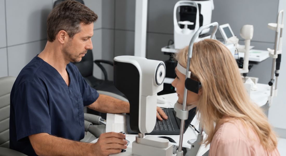
Your ophthalmologist may check your visual acuity, colour vision, pupils and eye movements to assess how well your optic nerves are working. Your visual field is also important because IIH can affect your peripheral vision before you notice a major change in your central vision.
Your optic nerves may be photographed and examined with OCT to monitor swelling and changes over time. Your results are considered together with your symptoms and clinical examination to understand how IIH is affecting your vision.
Evidence Note
Your visual function can be affected by IIH even when your central visual acuity remains relatively good. Formal visual-field testing is therefore important because peripheral visual loss can develop before you notice a major change in everyday central vision. OCT and optic-nerve photography can also help your ophthalmologist monitor papilloedema and structural changes over time.
Your monitoring results are considered together rather than relying on one test alone. Changes in visual fields, optic-nerve appearance, OCT findings and your symptoms can help your clinical team judge whether your IIH is stable or whether treatment needs to be adjusted to protect your vision.
Why Are Visual-Field Tests So Important?
Your visual-field test can detect changes in your peripheral vision even when your central vision still seems clear. Your brain may also compensate for gradual changes, so you may not notice them yourself.
Your ophthalmologist can compare your visual-field results over time to see whether your optic nerve function is stable or worsening. Your results can help guide treatment, particularly when papilloedema is active.
Research Insight
UK consensus guidance emphasises that protecting vision is one of the main principles of IIH management. The guidance recommends regular ophthalmic assessment of visual function and papilloedema because deterioration in visual function can change the urgency and type of treatment required.
Research and clinical guidance also show that treatment decisions should be based on the degree and rate of visual deterioration rather than headache symptoms alone. When visual function is declining significantly, treatment may need to become more urgent, including surgical options in severe cases, while stable disease may be managed with weight management, medication and ongoing monitoring.
Why Do You Need Brain Imaging?
Papilloedema can suggest raised pressure inside your skull, but it does not show what is causing that pressure. Your doctors therefore use brain imaging to check for conditions such as a brain mass, hydrocephalus or other structural problems.
When papilloedema is being investigated, brain imaging should include CT or MR venography to exclude cerebral venous sinus thrombosis. Your scan findings can support your assessment, but your doctors need to exclude other causes before confirming IIH.
What Happens During a Lumbar Puncture?
Your lumbar puncture involves a fine needle being placed in your lower back to reach the cerebrospinal fluid surrounding your brain and spinal cord. Your doctor can measure your cerebrospinal fluid pressure and collect a sample to check for other conditions.
Your lumbar puncture is usually performed after appropriate brain imaging and clinical assessment have excluded reasons why the procedure should not be performed.
Clinical Tip
If you are having a lumbar puncture as part of your IIH assessment, ask your doctor what your opening cerebrospinal fluid pressure was and how the result fits with your other findings. The pressure measurement is only one part of the diagnostic process, so your symptoms, eye examination and brain imaging also need to be considered together.
You should also tell your clinical team if your vision changes before or after the procedure. A lumbar puncture can temporarily reduce pressure, but it is generally not used as a long-term treatment strategy for IIH because the pressure can rise again.
How Is IIH Treated?
IIH treatment aims to protect your vision, reduce raised pressure inside your skull and manage symptoms such as headaches. Your treatment plan depends on your optic nerve swelling, visual changes and other symptoms.
Your weight may be an important part of treatment when relevant to your IIH. Your doctor may also prescribe medicines such as acetazolamide, while worsening vision may require more urgent treatment to protect your optic nerves.
Why Can Weight Management Be Important?
Your weight and recent weight gain can be important factors in IIH. When weight is contributing to your condition, sustained weight loss can help reduce pressure and improve your symptoms and papilloedema.
Your medical team can help you follow a structured weight-management plan. If your vision is already at risk, your doctors may use other treatments as well rather than relying on weight loss alone.
What Medicines Are Used for IIH?
Acetazolamide may be prescribed to reduce cerebrospinal fluid production and lower your intracranial pressure. Your dose can be adjusted according to your response and any side effects, such as tingling, altered taste, tiredness or stomach symptoms.
Your doctor may also consider topiramate in selected cases, particularly when you have migraine-type headaches. Topiramate has important pregnancy risks, and if you could become pregnant, your specialist must follow the current UK MHRA pregnancy-prevention requirements before and during treatment. Your medicines are only part of your care, so your visual fields and papilloedema still need regular monitoring.
IIH Assessment and Treatment Options
| Assessment or treatment | Main purpose | When it may be used |
| Visual-field testing | Detects changes in peripheral visual function | During diagnosis and follow-up to monitor optic-nerve function |
| OCT and optic-nerve imaging | Monitors papilloedema and structural changes | During ophthalmic monitoring |
| MRI or CT brain imaging | Looks for structural causes of raised intracranial pressure | During diagnostic assessment |
| MRV or CTV | Assesses the cerebral venous system and helps exclude venous thrombosis | When evaluating papilloedema or suspected raised intracranial pressure |
| Lumbar puncture | Measures cerebrospinal fluid opening pressure and analyses fluid | After appropriate imaging as part of confirming suspected IIH |
| Weight management | Addresses an important modifiable factor in IIH | Particularly when excess weight or recent weight gain is relevant |
| Acetazolamide | Reduces cerebrospinal fluid production | Commonly used when medication is appropriate, particularly when vision is affected |
| Topiramate | May help with headaches and has carbonic-anhydrase-inhibiting effects | Considered in selected patients |
| CSF diversion surgery | Diverts cerebrospinal fluid to reduce pressure | Considered when vision is threatened or deteriorating |
| Venous sinus stenting | Addresses suitable cerebral venous sinus narrowing | Considered in selected patients with appropriate venous sinus findings |
| Optic nerve sheath fenestration | Reduces pressure around the optic nerve | Considered in selected cases with threatened vision |
When Might Surgery Be Needed?

IIH will not usually require surgery, but your treatment may need to become urgent if your vision is worsening or your optic nerves are at serious risk. Your doctors may act quickly in severe or rapidly progressing cases to help prevent permanent sight loss.
Your specialist may consider procedures such as a shunt to divert cerebrospinal fluid or optic nerve sheath fenestration to reduce pressure around your optic nerve. Your specialist may also discuss venous sinus stenting in selected cases when your findings are suitable. The choice depends on your visual findings, anatomy, overall symptoms, local expertise and the balance of potential benefits and complications.
Myth vs Fact
| Myth | Fact |
| If your central vision is clear, IIH is not damaging your sight. | Peripheral visual loss can develop while central visual acuity remains relatively good, so formal visual-field testing is important. |
| Severe headaches always mean your optic nerves are at greater risk. | Headache severity does not reliably show how much visual damage is occurring. Your visual function needs to be assessed separately. |
| A lumbar puncture permanently treats IIH. | A lumbar puncture can temporarily reduce cerebrospinal fluid pressure, but it is generally not used as the main long-term treatment. |
| Everyone with IIH needs surgery. | Many people can be managed with weight management, medication and monitoring. Surgery is mainly considered when vision is threatened or deteriorating, particularly when deterioration is rapid or severe, or when other treatment is insufficient. |
| IIH only affects your eyesight temporarily. | Persistent raised pressure can cause optic-nerve damage and permanent vision loss if it is not controlled. |
Key Takeaways
- IIH causes raised pressure around the brain without another clear underlying cause.
- Papilloedema occurs when raised pressure causes swelling of the optic nerve heads.
- IIH can affect peripheral vision before you notice a major change in central vision.
- Visual-field testing is an important part of monitoring your optic-nerve function.
- OCT and optic-nerve photographs can help monitor papilloedema and structural changes.
- Brain imaging, including CT or MR venography during the investigation of papilloedema, helps exclude other causes of raised intracranial pressure, including cerebral venous sinus thrombosis.
- Lumbar puncture can measure cerebrospinal fluid pressure and help support the diagnosis.
- Weight management is an important part of treatment when relevant to your IIH.
- Acetazolamide may be used to reduce cerebrospinal fluid production and help protect visual function.
- Surgery may be needed urgently when your visual function is deteriorating or your optic nerves are at serious risk.
Frequently Asked Questions
- What is idiopathic intracranial hypertension (IIH)?
Idiopathic intracranial hypertension is raised pressure inside your skull without another clear cause. Before diagnosing IIH, your doctors need to exclude conditions such as a brain tumour, infection or blood clot. - How can IIH affect your vision?
Your raised intracranial pressure can cause swelling of your optic nerves, known as papilloedema. You may experience blurred vision, temporary dimming or loss of vision, reduced peripheral vision or double vision. - What is papilloedema?
Papilloedema is swelling of your optic nerve head caused by raised pressure inside your skull. Your ophthalmologist can identify the swelling during an eye examination and monitor whether it is affecting your optic nerve function. - Can IIH cause permanent vision loss?
Yes. If raised pressure continues to damage your optic nerves, it can cause permanent vision loss. Your central vision may initially remain clear even when your peripheral vision is starting to deteriorate. - What eye tests are used to monitor IIH?
Your ophthalmologist may check your visual acuity, colour vision, pupils and eye movements, along with formal visual-field testing. They may also photograph your optic nerves and use OCT to monitor swelling and changes over time. - Why are visual-field tests important in IIH?
Visual-field testing can identify changes in your peripheral vision before you notice a significant change in your central vision. Comparing your results over time helps your ophthalmologist determine whether your optic nerve function is stable or worsening. - How is IIH diagnosed?
Your diagnosis is based on your symptoms, eye and neurological examinations, appropriate brain and venous imaging and, after imaging, a lumbar puncture to measure cerebrospinal fluid opening pressure and analyse the fluid. Other causes of raised intracranial pressure need to be excluded first. - What happens during a lumbar puncture for IIH?
A lumbar puncture uses a fine needle in your lower back to measure your cerebrospinal fluid pressure and collect a sample for testing. It may temporarily lower your pressure, but repeated lumbar punctures are not usually used as long-term treatment. - How is idiopathic intracranial hypertension treated?
Your treatment may include weight management when relevant and medicines such as acetazolamide to reduce cerebrospinal fluid production. Topiramate may also be considered in selected cases, while worsening vision may require more urgent treatment. - When might surgery be needed for IIH?
Surgery may be considered when your vision is worsening or your optic nerves are at serious risk. Depending on your individual findings, options may include a shunt, optic nerve sheath fenestration or, in selected cases, venous sinus stenting.
Final Thoughts: IIH and Vision
Idiopathic intracranial hypertension can affect your optic nerves and vision, particularly when raised pressure causes persistent papilloedema. Regular monitoring of your visual fields, optic nerves and overall eye health is important because early changes may not always be obvious to you. If your vision is worsening, prompt assessment can help reduce the risk of permanent sight loss.
If you would like to discuss your symptoms with an experienced eye specialist, you can contact the Eye Clinic London team for a comprehensive assessment.
References:
- Toro, M.D., Castellino, N., Russo, A., Scollo, D., Avitabile, T., Rejdak, R., Rejdak, M., Cimino, V., Costagliola, C., Carnevali, A. and Chisari, C.G. (2024) ‘Optic Nerve Head and Retinal Changes in Idiopathic Intracranial Hypertension: Correlation with Short-Term Cerebrospinal Fluid Pressure Monitoring’, Journal of Clinical Medicine, 13(2), 562. Available at: https://www.mdpi.com/2077-0383/13/2/562
- Mollan, S.P., Davies, B., Silver, N.C., Shaw, S., Mallucci, C.L., Wakerley, B.R., Krishnan, A., Chavda, S.V., Ramalingam, S., Edwards, J., Hemmings, K., Williamson, M., Burdon, M.A., Hassan-Smith, G., Digre, K., Liu, G.T., Jensen, R.H. and Sinclair, A.J. (2018) ‘Idiopathic intracranial hypertension: consensus guidelines on management’, Journal of Neurology, Neurosurgery & Psychiatry, 89(10), pp. 1088–1100. Available at: https://pubmed.ncbi.nlm.nih.gov/29903905/
- The NORDIC Idiopathic Intracranial Hypertension Study Group Writing Committee (2014) ‘Effect of acetazolamide on visual function in patients with idiopathic intracranial hypertension and mild visual loss: the Idiopathic Intracranial Hypertension Treatment Trial’, JAMA, 311(16), pp. 1641–1651. Available at: https://pubmed.ncbi.nlm.nih.gov/24756514/
- Wakerley, B.R., Mollan, S.P. and Sinclair, A.J. (2020) ‘Idiopathic intracranial hypertension: Update on diagnosis and management’, Clinical Medicine, 20(4), pp. 384–388. Available at: https://pmc.ncbi.nlm.nih.gov/articles/PMC7385768/
- Raoof, N. and Hoffmann, J. (2021) ‘Diagnosis and treatment of idiopathic intracranial hypertension’, Cephalalgia, 41(4), pp. 472–478. Available at: https://pmc.ncbi.nlm.nih.gov/articles/PMC8020303/