


The History of Artificial Intelligence in Ophthalmology
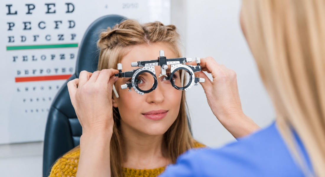
Artificial intelligence may seem like a recent development in eye care, but researchers have explored computer-assisted diagnosis in ophthalmology for several decades. If you have an eye examination today, you may encounter digital imaging and computer-based analysis that can help your specialist identify and monitor eye conditions.
As technology has advanced, AI has become increasingly capable of analysing retinal photographs, optical coherence tomography scans and other clinical information. For you, these developments may support image interpretation, screening or earlier identification of certain abnormalities when the AI system has been appropriately validated for that specific task.
What Does Artificial Intelligence Mean in Ophthalmology?
Artificial intelligence (AI) refers to computer technologies that can recognise patterns, analyse information and make predictions. In ophthalmology, AI can help analyse your retinal photographs, measure structures in your OCT scans or identify patterns that may need further specialist assessment.
You may also hear terms such as machine learning and deep learning. Machine-learning systems identify statistical patterns from training data, while deep learning uses multilayer neural networks that can learn increasingly complex representations from examples such as retinal photographs or OCT scans.
Key Milestones in AI and Ophthalmology
| Period/Year | Milestone | Why it mattered |
| Mid-1970s | CASNET/Glaucoma developed | One of the earliest knowledge-based computer consultation systems applied to ophthalmology |
| 1976 | CASNET/Glaucoma publicly evaluated | Demonstrated computer-assisted glaucoma consultation alongside specialist opinion |
| 1978 | Glaucoma Consultation by Computer published | Documented an early ophthalmic AI consultation system |
| 1991 | Landmark OCT paper published | Demonstrated non-invasive cross-sectional optical imaging and helped establish a technology that would later become fundamental to ophthalmic imaging and AI research. |
| 2010s | Deep learning expands | Enabled algorithms to learn complex image features directly from large datasets |
| 2016 | Gulshan diabetic-retinopathy study | Major demonstration of deep learning on retinal photographs |
| 2017 | Ting multiethnic validation | Tested AI across a very large, diverse retinal-image dataset |
| 2018 | Autonomous DR trial | Tested autonomous AI prospectively in primary care |
| 2018 | FDA authorises IDx-DR | Major regulatory milestone for autonomous medical AI |
| 2018 | Moorfields–DeepMind OCT study | Demonstrated AI referral recommendations across retinal diseases |
The Early Computer Era in Ophthalmology
Some of the earliest links between artificial intelligence and ophthalmology appeared during the 1970s. Although computers were far less powerful than the technology you use today, researchers were already exploring whether computers could organise medical knowledge and support clinical reasoning.
Glaucoma became an important area of early research because your diagnosis and treatment can involve several interacting factors. One notable project was CASNET (Causal Associational Network), which represented disease processes as linked networks to help your specialist consider glaucoma diagnosis, prognosis and treatment decisions.
CASNET and the First Glaucoma AI Systems
CASNET/Glaucoma was an important milestone in the early history of artificial intelligence in eye care. Developed and demonstrated during the 1970s, it used stored specialist knowledge to interpret clinical findings, model glaucoma pathophysiology and support diagnostic, prognostic and management recommendations.
The system was very different from the AI you may encounter today because it did not learn from large collections of eye images. Instead, it relied on organised medical knowledge and relationships between disease states to process information and provide useful support to your doctor when making clinical decisions.
Why Ophthalmology Became Well Suited to AI
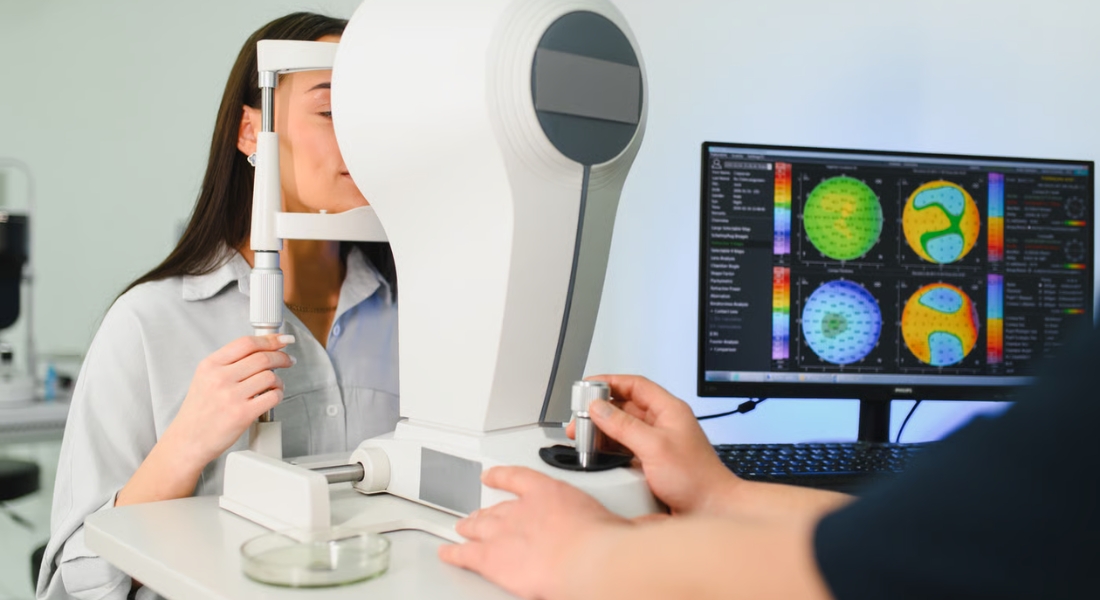
Ophthalmology is particularly suitable for artificial intelligence because much of your eye assessment relies on digital images and measurements. Your retinal photographs, OCT scans, optic nerve images and corneal maps can be stored digitally and analysed by computer systems.
The widespread use of digital imaging provides large datasets that can be used to develop and validate image-analysis algorithms. However, a large dataset alone does not guarantee a reliable AI system. Image quality, annotation quality, patient diversity and external validation all affect performance. By studying many eye images, an algorithm can recognise patterns linked to certain abnormalities and potentially support your specialist’s assessment, although its accuracy still depends on the quality of the images and data used for training.
The Era of Rule-Based Expert Systems
Early artificial intelligence relied heavily on rules and specialist knowledge entered into a computer. Your symptoms, test results and other clinical findings could be processed through these predefined rules to produce a possible recommendation, but the system could only work within the information it had been given.
This approach was useful but could not fully reproduce your ophthalmologist’s clinical judgement. Your doctor may consider imaging, examination findings, symptoms and your previous eye history together, so researchers began developing systems that could learn patterns from data rather than depending entirely on manually programmed rules.
OCT Created a New Source of Ophthalmic Data
Optical coherence tomography (OCT) created a major new source of detailed digital information for ophthalmology. If you have an OCT scan, it can produce cross-sectional images of your retina and optic nerve that allow your specialist to assess retinal structures in considerable detail and provide data that appropriately trained AI systems can analyse.
- Detailed Retinal Imaging: OCT can show individual retinal layers and structural changes that may not be visible on a standard retinal photograph.
- Objective Measurements: Your scans can provide measurable information about retinal thickness, fluid and other structural features.
- Monitoring Over Time: Comparing OCT scans from different appointments can help identify changes in your eye condition or response to treatment.
- Supporting AI Analysis: Large collections of OCT scans have allowed researchers to train AI systems to recognise and classify patterns associated with retinal disease.
OCT therefore became an important foundation for modern ophthalmic AI as well as routine clinical care. Its detailed and measurable imaging data can support diagnosis and monitoring, while your ophthalmologist remains responsible for interpreting the results in the context of your overall eye health.
Machine Learning Marked an Important Turning Point
Machine learning changed how researchers approached computer-assisted eye care by allowing systems to learn patterns from examples rather than relying entirely on manually written rules. If you provide an algorithm with thousands of retinal photographs that have already been assessed by specialists, it can learn features that help distinguish between different conditions.
This did not mean that computers understood your eye disease in the same way your ophthalmologist does. Their performance still depends on the quality of the training data, the accuracy of the clinical labels and whether the people represented in the dataset are similar to the patients who will eventually use the system.
Deep Learning Accelerated Progress in the 2010s
Progress in ophthalmic AI accelerated during the 2010s as deep learning, larger image datasets and more powerful computers became available. If you have a retinal photograph or OCT scan, appropriately trained deep-learning systems can learn complex image features from training examples without researchers manually specifying every relevant feature beforehand.
Convolutional neural networks became particularly useful for analysing these detailed images and identifying subtle changes. This development gave your ophthalmologist new ways to assess your eye health, while allowing you to benefit from AI tools that could perform specific image-analysis tasks more efficiently.
Digital Retinal Photography Changed What Computers Could See
The growing use of digital retinal photography created an important foundation for modern ophthalmic AI. If you have a digital photograph of the back of your eye, it can show detailed information about your retina, optic disc and blood vessels that computers can analyse.
Digital images also allowed researchers to build larger datasets and train machine-learning systems to recognise patterns linked to particular diseases. This was a major change from early expert systems because computers could analyse your eye image directly, helping pave the way for important AI developments such as diabetic retinopathy screening.
Diabetic Retinopathy Became a Landmark Application
Diabetic retinopathy became one of the most important early applications of deep learning in ophthalmology. If you have diabetes, changes in the small blood vessels of your retina can develop gradually, so regular retinal screening can help identify problems before they become more advanced.
AI became particularly useful because screening programmes may need to assess large numbers of retinal photographs. A landmark 2016 study showed that a deep-learning system could detect diabetic retinopathy with high sensitivity and specificity in the datasets studied, giving your screening team another potential tool for identifying images that need further attention.
Research Insight
Earlier computer systems depended heavily on manually programmed knowledge or features selected by researchers. The system was trained and evaluated for detecting referable diabetic retinopathy, which in the study included moderate-or-worse diabetic retinopathy, referable diabetic macular oedema, or both.
The result helped establish retinal photography as one of the leading medical-imaging applications for deep learning, but it did not by itself prove that the algorithm would perform identically in every population, camera system or clinical environment. Later external and prospective validation therefore became essential.
AI Began Moving Beyond Research Experiments
As AI systems began showing strong results in research studies, researchers needed to find out whether they could also work reliably with real patients. If you have an eye examination in everyday clinical practice, factors such as image quality, camera type and your background can differ from the carefully controlled conditions used during development.
Researchers therefore started testing AI across different populations and healthcare settings. A 2017 study developed and validated a deep-learning system using data comprising 494,661 retinal images across multiethnic populations with diabetes.
Autonomous AI Became Possible in Diabetic Eye Screening
Autonomous AI systems marked an important step in diabetic retinopathy screening because they can analyse your retinal photographs and provide a result for a specific authorised task. If you have diabetes, this could help identify signs of retinopathy without requiring a specialist to review every image first.
A pivotal trial involving 900 people with diabetes showed the potential of this approach in primary care. However, you still need a wider eye assessment when appropriate because autonomous AI is designed for specific screening tasks rather than replacing your specialist’s judgement.
AI Returned to Glaucoma With Modern Imaging
Glaucoma has come full circle in the history of ophthalmic AI. It was an early focus of computer-assisted clinical reasoning in the 1970s and remains an important area of research today. Modern research systems can analyse fundus photographs, OCT scans, visual-field data and optic-nerve measurements for patterns associated with glaucoma.
Modern research is also exploring whether AI can help predict your risk of developing or progressing glaucoma. This could eventually support more personalised monitoring, although these tools still need careful clinical validation before they can guide your individual care.
AI Expanded Into AMD and Other Retinal Conditions
Age-related macular degeneration (AMD) became another important area for AI research because retinal imaging plays a major role in diagnosis and monitoring. AI research systems can analyse retinal photographs or OCT scans for features associated with AMD, and some tools are being investigated for segmentation, disease-activity assessment and progression prediction.
AI research has expanded beyond diabetic retinopathy into glaucoma, AMD and a range of other imaging-based ophthalmic conditions. This means you may increasingly encounter AI tools that support your specialist in detecting changes, monitoring progression and understanding your eye health.
The Moorfields and DeepMind OCT Research
In 2018, researchers at Moorfields Eye Hospital and DeepMind reported a deep-learning system designed to analyse OCT scans and support referral decisions for retinal disease. If you have an OCT scan, this type of AI can assess detailed retinal information and help identify cases that may need further specialist attention.
This was important because AI was moving beyond simply deciding whether an image looked abnormal. For you, it showed how AI could potentially support referral and care pathways, helping your specialist identify which patients may need more urgent assessment or monitoring.
How Is AI Being Used and Studied in Modern Ophthalmology?
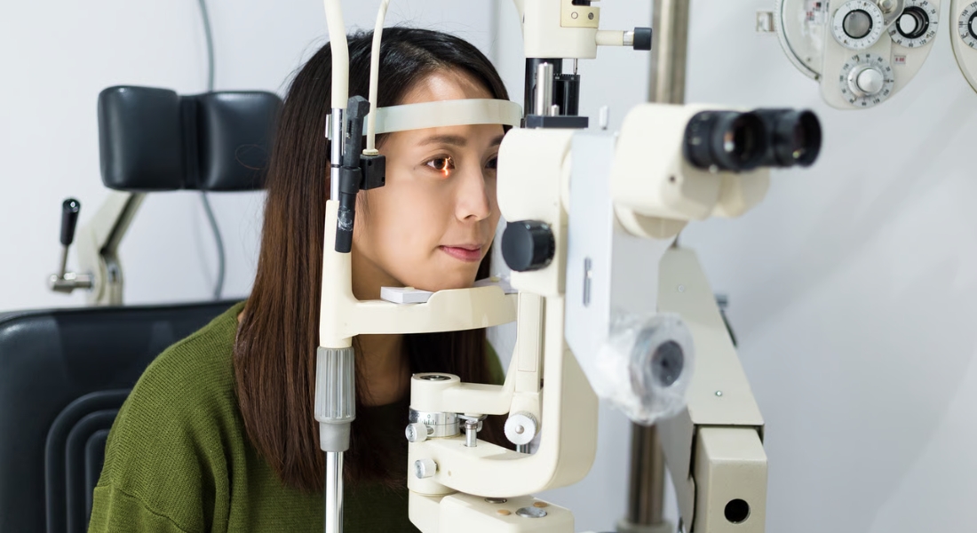
In some jurisdictions, AI systems have regulatory authorisation for specific screening tasks, while many other ophthalmic applications remain in research or clinical-development stages. Researchers are studying AI for image classification, segmentation, triage, disease monitoring and prediction using retinal photographs, OCT, visual fields and other clinical data.
Whether an AI output should influence care depends on the particular system, the task for which it was validated, image quality, the patient population and how the result fits with the wider clinical assessment. An algorithm that performs well for one imaging task should not automatically be assumed to perform equally well for another disease, device or population.
Myth vs Fact
| Myth | Fact |
| AI in ophthalmology only appeared in the last few years. | Computer-assisted glaucoma decision systems were already being developed during the 1970s. |
| Early ophthalmic AI analysed retinal photographs like modern deep learning. | Early systems such as CASNET primarily relied on encoded clinical knowledge and disease relationships. |
| Machine learning and deep learning are exactly the same thing. | Deep learning is a subset of machine learning that uses multilayer neural networks to learn complex patterns. |
| OCT has always existed alongside AI. | A landmark 1991 paper first described and demonstrated OCT imaging; the technology subsequently developed into a major clinical ophthalmic imaging platform. |
| The 2016 diabetic-retinopathy study meant AI was immediately ready for every clinic. | Strong retrospective performance still required external and prospective clinical validation. |
| Autonomous AI means a computer can manage all of your eye care. | Autonomous systems are authorised for defined tasks; broader assessment and treatment can still require clinicians. |
| The Moorfields–DeepMind study proved AI could replace retinal specialists. | It demonstrated strong referral performance on a defined OCT task, not replacement of full clinical care. |
| AI is automatically equally accurate for every population. | Performance can change if training data, patient populations, imaging devices or disease patterns differ. |
Key Takeaways
- Ophthalmic AI has roots in computer-assisted glaucoma consultation systems developed during the mid-1970s.
- CASNET/Glaucoma relied on encoded expert knowledge and causal disease models rather than modern image-based learning.
- A landmark 1991 paper demonstrated OCT, which later became a major source of digital ophthalmic data for both clinical care and AI research.
- Deep learning accelerated ophthalmic image analysis during the 2010s.
- The 2016 Gulshan study trained on 128,175 retinal photographs and demonstrated strong performance for referable diabetic-retinopathy detection in two validation datasets.
- The 2017 Ting study extended validation across a very large multiethnic collection of retinal images.
- A prospective 2018 primary-care study of 900 people demonstrated the feasibility of autonomous AI diabetic-retinopathy screening.
- The FDA granted De Novo classification to IDx-DR on 11 April 2018 for a defined diabetic-retinopathy detection task.
- The 2018 Moorfields–DeepMind study trained on 14,884 OCT scans and achieved referral performance reaching or exceeding experts on its defined retinal pathway.
Frequently Asked Questions
- When did artificial intelligence first become part of ophthalmology?
Early computer-assisted approaches to ophthalmology were being explored during the 1970s. Systems such as CASNET showed that computers could use organised medical knowledge to support clinical decision-making, particularly in areas such as glaucoma. - Why is ophthalmology well suited to artificial intelligence?
Ophthalmology relies heavily on digital images and measurable structures, including retinal photographs, OCT scans and optic nerve images. This provides AI systems with large amounts of visual data that can be analysed for patterns associated with eye conditions. - How did retinal photography help develop ophthalmic AI?
Digital retinal photography allowed researchers to collect and analyse large numbers of standardised eye images. This helped machine-learning systems learn patterns associated with conditions such as diabetic retinopathy. - When did machine learning become important in ophthalmology?
Machine learning became increasingly important as researchers gained access to larger datasets and more powerful computers. During the 2010s, deep-learning techniques significantly accelerated research into automated analysis of retinal photographs and other ophthalmic images. - What was one of the first major uses of AI in eye care?
Diabetic retinopathy screening became one of the most significant early applications of deep learning in ophthalmology. AI can analyse retinal photographs and identify changes that may require further assessment, helping screening programmes manage large numbers of images. - Can AI detect glaucoma?
AI can analyse retinal photographs, OCT scans, visual fields and optic nerve measurements to identify patterns associated with glaucoma. Researchers are also investigating whether AI can help predict disease progression, although your ophthalmologist still needs to interpret these findings alongside your wider clinical information. - How has OCT contributed to the development of AI?
OCT produces detailed cross-sectional images of your retina and optic nerve, creating large amounts of structured imaging data for AI systems to analyse. This has helped researchers develop tools that can identify retinal abnormalities and support diagnosis, monitoring and referral decisions. - Can AI replace an ophthalmologist?
No. AI is designed to support specific tasks such as image analysis, screening or monitoring, while your ophthalmologist considers your symptoms, medical history, examination findings and treatment response when making decisions about your care. - How is AI being used for age-related macular degeneration?
AI can analyse retinal photographs and OCT scans to identify and monitor changes associated with age-related macular degeneration. This may help your ophthalmologist assess disease activity, monitor progression and support treatment planning. - What does the future of AI in ophthalmology look like?
AI is likely to become increasingly useful for earlier detection, personalised monitoring, imaging analysis and treatment planning across a range of eye conditions. As these systems develop, careful clinical validation, diverse training data and appropriate oversight will remain important to ensure they provide safe and reliable support for your eye care.
Final Thoughts: The Future of AI in Eye Care
Artificial intelligence has come a long way in ophthalmology, from early computer-assisted systems to advanced tools that can analyse retinal photographs and OCT scans. For you, these developments may support earlier detection, more detailed monitoring and more personalised eye care, while your ophthalmologist continues to interpret the findings and make decisions about your treatment.
As AI continues to develop, careful clinical validation and appropriate oversight will remain important for safe and reliable use. If you would like to learn more about how advances in technology may support your eye health, you can speak with one of our specialists at Eye Clinic London about your individual needs and the options that may be appropriate for you.
References:
- Weiss, S.M., Kulikowski, C.A. and Safir, A. (1978) ‘Glaucoma consultation by computer’, Computers in Biology and Medicine, 8(1), pp. 25–40. Available at: https://www.sciencedirect.com/science/article/pii/0010482578900112
- Huang, D., Swanson, E.A., Lin, C.P., Schuman, J.S., Stinson, W.G., Chang, W., Hee, M.R., Flotte, T., Gregory, K., Puliafito, C.A. and Fujimoto, J.G. (1991) ‘Optical coherence tomography’, Science, 254(5035), pp. 1178–1181. Available at: https://pmc.ncbi.nlm.nih.gov/articles/PMC4638169/
- Gulshan, V., Peng, L., Coram, M., Stumpe, M.C., Wu, D., Narayanaswamy, A., Venugopalan, S., Widner, K., Madams, T., Cuadros, J. et al. (2016) ‘Development and validation of a deep learning algorithm for detection of diabetic retinopathy in retinal fundus photographs’, JAMA, 316(22), pp. 2402–2410. Available at: https://pubmed.ncbi.nlm.nih.gov/27898976/
- Abràmoff, M.D., Lavin, P.T., Birch, M., Shah, N. and Folk, J.C. (2018) ‘Pivotal trial of an autonomous AI-based diagnostic system for detection of diabetic retinopathy in primary care offices’, npj Digital Medicine, 1, article 39. Available at: https://pmc.ncbi.nlm.nih.gov/articles/PMC6550188/
- De Fauw, J., Ledsam, J.R., Romera-Paredes, B., Nikolov, S., Tomasev, N., Blackwell, S., Askham, H., Glorot, X., O’Donoghue, B., Visentin, D. et al. (2018) ‘Clinically applicable deep learning for diagnosis and referral in retinal disease’, Nature Medicine, 24(9), pp. 1342–1350. Available at: https://pubmed.ncbi.nlm.nih.gov/30104768/