


Why Did Sugar Ray Leonard Need Eye Surgery?
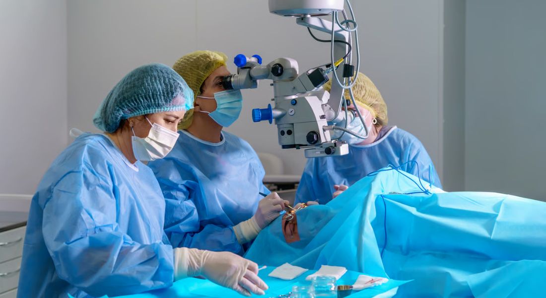
Sugar Ray Leonard needed eye surgery in 1982 after doctors found a partially detached retina in his left eye. He developed blurred vision and spots in his left eye while preparing for a title defence, so the fight was cancelled and he underwent a two-hour operation at the Wilmer Eye Institute.
His case shows why eye injuries can be serious, particularly in boxing. His surgeon could not confirm exactly when or how the retinal injury happened, so it would be wrong to blame one specific punch. If you notice new flashes, floaters or a curtain-like shadow across your vision, you should seek urgent eye care.
Why Did Sugar Ray Leonard Have Eye Surgery?
In 1982, Sugar Ray Leonard developed blurred vision and spots while training for a title defence. Doctors found a partially detached retina in his left eye, his fight was cancelled and he was treated at the Wilmer Eye Institute.
Surgery aimed to reattach the retina and protect his sight. Sudden flashes, new floaters, blurred vision or a curtain-like shadow need urgent assessment because retinal detachment can permanently affect vision.
What Is the Retina and Why Is It So Important?
Your retina is a thin layer of light-sensitive tissue at the back of your eye. You need it to turn light into signals that travel through your optic nerve to your brain, allowing you to see. If your retina is damaged or detached, glasses cannot correct the problem because the issue is not simply with focus.
In a retinal detachment, the light-sensitive neurosensory retina separates from the underlying retinal pigment epithelium. This disrupts normal retinal function and can lead to permanent sight loss if an important area remains detached. Your macula provides your sharp central vision, so if a detachment reaches it, your central vision can become significantly worse.
What Is a Retinal Detachment?

A retinal detachment happens when your retina separates from the tissue underneath it that normally supports and nourishes it. In many cases, a tear or hole allows fluid to pass beneath the retina, lifting it away from the back of your eye and affecting your vision.
This type is called a rhegmatogenous retinal detachment, although other types can develop because of scar tissue or fluid build-up. A detached retina is usually painless, so you may not realise how serious the problem is. If you notice sudden flashes, new floaters or a shadow across your vision, you should seek urgent eye care to reduce the risk of permanent sight loss.
How Can Boxing or Eye Trauma Cause Retinal Detachment?
A forceful blow to your eye can suddenly compress and distort your eyeball, putting stress on the retina and the vitreous gel inside it. This can sometimes cause a retinal tear, allowing fluid to pass underneath the retina and lead to detachment. A severe direct blow to your eye can increase your risk of retinal detachment.
If you take part in boxing or another contact sport, repeated impacts can be particularly relevant. You may not notice a serious problem immediately after an injury, so if you develop new flashes, floaters, blurred vision or a shadow across your vision, you should have your eyes assessed urgently.
Did One Particular Punch Cause Leonard’s Detached Retina?
There is no reliable evidence that one specific punch caused Sugar Ray Leonard’s retinal detachment. His ophthalmologist said at the time that it was impossible to determine exactly when or how the damage occurred, although previous injuries to his left eye and repeated boxing trauma were considered possible factors.
Leonard had suffered significant swelling around his left eye during his 1981 fight with Thomas Hearns and had also been sparring while preparing for his next fight. However, you should not assume that either event directly caused the detachment. The safest conclusion is that significant eye trauma can increase your risk, but the exact cause of Leonard’s injury was never established.
What Warning Signs Can a Retinal Detachment Cause?
A retinal detachment can cause sudden changes in your vision, often without pain. Recognising these warning signs early is important.
- New floaters: You may suddenly notice dots, threads or cobweb-like shapes in your vision.
- Flashes of light: Brief flashes may appear, particularly towards the edge of your sight.
- Blurred vision: Your vision may become suddenly or increasingly unclear.
- Dark shadow: A curtain-like shadow may spread across part of your vision.
These symptoms do not always mean your retina has detached, but they need urgent assessment. Prompt treatment can help reduce the risk of permanent sight loss.
Who Else Is at Risk of Retinal Detachment?
You do not have to be a boxer or have an eye injury to develop a retinal detachment. As you get older, the vitreous gel inside your eye naturally changes and can pull away from the retina. This is called posterior vitreous detachment (PVD) and, although most PVDs do not cause retinal detachment, they can sometimes lead to a retinal tear.
You may also have a higher risk if you are very short-sighted, have previously had cataract surgery, have had a retinal detachment in your other eye, have a family history of retinal detachment or have certain inherited retinal conditions. Previous significant eye trauma can also increase your risk.
How Is a Retinal Detachment Diagnosed?
Your ophthalmologist will examine the back of your eye, usually after using drops to dilate your pupil. You may have temporary blurred vision and light sensitivity afterwards, so you should follow your clinic’s advice about driving. Your specialist will check for retinal tears, holes or areas where the retina has detached.
If the retina cannot be seen clearly, an ocular ultrasound may sometimes be used to help assess whether a detachment is present. If a retinal tear is found before the retina detaches, treatment may prevent further progression. Once a significant detachment has developed, you will usually need surgery to reattach the retina.
What Is the Difference Between a Retinal Tear and a Retinal Detachment?
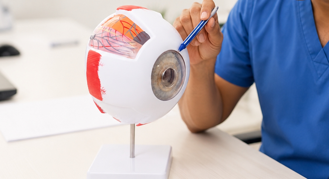
A retinal tear is a break in your retina, while a retinal detachment happens when fluid passes through the tear and lifts the retina away from the back of your eye. You can therefore have a retinal tear without having a full detachment, which gives your ophthalmologist an opportunity to treat it before the problem progresses.
Laser treatment or cryotherapy can sometimes seal a retinal tear and reduce the risk of detachment. However, once your retina has detached significantly, you will usually need surgery to put it back into position. Because you cannot tell the difference from symptoms alone, new flashes or floaters should be assessed promptly.
Retinal Tear vs Retinal Detachment
| Feature | Retinal Tear | Retinal Detachment |
| What happens | A break or hole develops in the retina | The retina separates from the supporting tissue at the back of the eye |
| How it may develop | Often occurs when the vitreous pulls on the retina or after eye trauma | Fluid may pass through a retinal tear and collect underneath the retina |
| Possible symptoms | New flashes, sudden floaters or other visual changes | Flashes, floaters, blurred vision or a dark curtain or shadow |
| Pain | Usually painless | Usually painless |
| Treatment | Laser or cryotherapy may sometimes be used to seal the tear | Usually requires retinal surgery to reposition and support the retina |
| Why early assessment matters | Treatment may prevent progression to detachment | Prompt treatment can reduce the risk of permanent sight loss |
| Effect on vision | Vision may remain relatively unaffected if treated before detachment | Vision can be significantly reduced, particularly if the macula detaches |
What Surgery Did Sugar Ray Leonard Have?
Sugar Ray Leonard had retinal surgery on his left eye at the Wilmer Eye Institute at Johns Hopkins University on 9 May 1982. Contemporary reports described cryotherapy around the damaged retinal area together with silicone material used to support the outer wall of the eye. This is consistent with the principles of scleral-buckle surgery, although the historical reports do not provide the same operative detail that would be recorded for a modern retinal procedure.
Your visual recovery after retinal surgery can depend on how long your retina was detached, whether your macula was affected and whether any retinal cells were permanently damaged. Leonard eventually recovered well, with reports in October 1982 stating that his vision had returned to normal and that his ophthalmologist had cleared him to decide whether he wanted to box again.
How Is a Detached Retina Treated Today?
Modern retinal-detachment surgery aims to close the retinal tear and put your retina back against the supporting tissue underneath it. The treatment your retinal surgeon recommends will depend on the location and extent of the detachment, the condition of your vitreous and the health of your eye.
You may have a vitrectomy, where the vitreous gel is removed and the retinal breaks are treated. A gas bubble or, in selected cases, silicone oil may then be used to support the retina while it heals. Another option is a scleral buckle, which supports the retina from outside the eye. In selected cases, pneumatic retinopexy may be used, with a gas bubble helping to reposition the retina.
What Happens During a Vitrectomy?
During a vitrectomy, your surgeon removes the vitreous gel from inside your eye using very small instruments. This reduces the pulling force on your retina, allowing the surgeon to flatten the detached retina and treat the tear with laser or freezing treatment.
Your surgeon may place a gas bubble inside your eye to hold the retina in position while it heals, and you may need to keep your head in a particular position afterwards. If you have an intraocular gas bubble, you must not fly or travel to high altitude until your ophthalmologist confirms the gas has completely disappeared. You should also tell any doctor, dentist or anaesthetist that gas is present in your eye because nitrous oxide must be avoided while the bubble remains.
What Is a Scleral Buckle and Why Is Cryotherapy Used?
A scleral buckle supports your retina from outside the eye. Your surgeon places a silicone band or piece of material around the white outer wall of your eye, gently pushing it inwards to reduce the pulling forces around the retinal tear and help the retina stay in place while it heals.
Cryotherapy uses controlled freezing around a retinal tear to create scar tissue that helps seal the break. Laser treatment can achieve a similar effect using controlled burns. Your retinal surgeon will choose the most suitable approach based on the location and type of retinal detachment. Leonard’s 1982 surgery was also reported to involve freezing treatment and silicone support.
Will Your Vision Return After Retinal Detachment Surgery?
Retinal-detachment surgery aims to reattach your retina and prevent further sight loss, but it cannot guarantee that your vision will return to how it was before. Your outcome depends on factors such as whether your macula was affected and how long the retina remained detached. If your macula detached, your central vision may improve but can remain blurred or reduced.
Your vision may also be very blurred immediately after surgery and can take several weeks to improve. If you have a gas bubble in your eye, your sight may remain limited until it gradually disappears. Leonard’s recovery was reportedly good, and he was later medically cleared to decide whether he wanted to return to boxing.
Evidence Note
A 2022 meta-analysis of 20 observational studies involving 1,929 patients found that macula-off rhegmatogenous retinal detachments repaired within 0–3 days of symptom onset had better final visual acuity than those repaired within 4–7 days. It also found better final visual acuity when macula-on detachments were repaired within 24 hours of presentation rather than after 24 hours.
These findings do not mean that every retinal detachment has the same surgical deadline. The evidence was of moderate quality for the macula-off comparison and lower quality for the macula-on comparison, while urgency also depends on the location and extent of the detachment and the retinal surgeon’s assessment. The important patient message is to seek urgent assessment rather than trying to judge the timing yourself.
When Should You Seek Urgent Assessment for Retinal Symptoms?
If you suddenly develop flashes for the first time, a sudden increase in floaters, blurred vision or a dark curtain or shadow across your sight, arrange urgent eye assessment. In the UK, request an urgent optometrist appointment or contact NHS 111 for advice about where you should be assessed.
You should not wait for pain because retinal detachment is usually painless. If you are very short-sighted, have had cataract or retinal surgery, or recently suffered an eye injury, tell your eye specialist about your history. Prompt assessment can help identify a retinal tear or detachment before permanent sight loss occurs.
Myth vs Fact
| Myth | Fact |
| A retinal detachment always causes eye pain. | No. Retinal detachment is usually painless, which is why sudden visual symptoms should not be ignored. |
| Every new floater means your retina is detached. | No. Floaters are common, but sudden new or increasing floaters especially with flashes, blur or a shadow need urgent assessment. |
| One specific punch definitely caused Sugar Ray Leonard’s retinal detachment. | No. His surgeon said it was impossible to establish exactly when the retinal injury occurred, although boxing trauma was a possible contributor. |
| A retinal tear and a detached retina are the same thing. | No. A tear is a break in the retina; detachment occurs when fluid passes underneath and lifts the retina away from the supporting tissue. |
| Successful surgery guarantees normal vision. | No. The retina can be anatomically reattached while some visual loss remains, particularly if the macula was detached or retinal tissue was damaged. |
| You can fly while a gas bubble remains in your eye after retinal surgery. | No. Reduced atmospheric pressure can cause an intraocular gas bubble to expand dangerously. Nitrous oxide must also be avoided until the bubble has completely disappeared. |
Key Takeaways
- Sugar Ray Leonard underwent surgery for a partially detached retina in his left eye on 9 May 1982 at the Wilmer Eye Institute.
- His surgeon could not establish exactly when or how the retinal injury occurred, so it should not be attributed to one particular punch.
- Retinal detachment is usually painless but can cause sudden floaters, flashes, blurred vision or a curtain-like shadow.
- Blunt eye trauma, including contact-sport injuries, can increase the risk of retinal tears and traumatic retinal detachment.
- A retinal tear can sometimes be treated before a significant detachment develops.
- Modern retinal-detachment repair may involve vitrectomy, scleral buckling or, in selected cases, pneumatic retinopexy.
- Visual recovery depends strongly on factors including whether the macula was involved and how long it had been detached.
- If gas is placed inside the eye, you must avoid flying until it has disappeared and tell healthcare professionals before receiving nitrous oxide.
- Sudden new flashes, floaters, blurred vision or a shadow/curtain require urgent assessment, even if your eye does not hurt.
Frequently Asked Questions
- Why did Sugar Ray Leonard need eye surgery?
Sugar Ray Leonard needed surgery in 1982 after doctors found a partially detached retina in his left eye. He had blurred vision while training for a title defence, leading to his fight being cancelled. - What happened to Sugar Ray Leonard’s retina?
Part of Leonard’s left retina had become detached, affecting his vision. His doctors treated the problem surgically to reattach the retina and protect his remaining sight. - Can boxing cause retinal detachment?
A strong blow to the eye can damage the retina and increase your risk of a retinal tear or detachment. If you develop flashes, new floaters or blurred vision after an injury, you should seek urgent assessment. - What are the warning signs of retinal detachment?
You may notice sudden flashes of light, a sudden increase in floaters, blurred vision or a dark curtain or shadow across your sight. These symptoms need urgent assessment because retinal detachment can cause permanent sight loss. - Is retinal detachment painful?
Usually, no. You can have a retinal detachment without experiencing pain, which is why sudden changes in your vision should not be ignored. - How is retinal detachment diagnosed?
Your ophthalmologist will usually examine the back of your eye after dilating your pupil. They will check for retinal tears, holes and areas where the retina has detached. - How is retinal detachment treated?
Treatment depends on the location, extent and type of retinal detachment. Repair may involve vitrectomy, scleral buckling or, in selected cases, pneumatic retinopexy. Laser treatment or cryotherapy is used to seal retinal tears as part of the treatment. - Can vision return after retinal detachment surgery?
Your vision may improve after surgery, but recovery depends on factors such as whether your macula was affected and how long the retina was detached. Surgery mainly aims to reattach the retina and prevent further sight loss. - Can a retinal tear become a retinal detachment?
Yes. Fluid can pass through a retinal tear and collect underneath the retina, causing it to detach. Treating a retinal tear early can sometimes prevent a retinal detachment from developing. - When should you seek urgent eye assessment?
Arrange urgent assessment if you suddenly develop new or increasing floaters, flashes, blurred vision or a dark curtain or shadow across your sight. In the UK, request an urgent optometrist appointment or contact NHS 111 for advice about where you should be assessed. Do not wait for pain because retinal detachment is usually painless.
Final Thoughts: Protecting Your Vision From Retinal Detachment
Sugar Ray Leonard’s experience highlights why changes in your vision should never be ignored, particularly after an eye injury or contact sport. Retinal detachment is usually painless, but symptoms such as sudden flashes, new floaters, blurred vision or a dark curtain across your sight can indicate a serious problem that needs urgent assessment. Acting quickly can help protect your remaining vision.
If you have concerns about your eyesight, our team at Eye Clinic London can provide a comprehensive assessment and discuss whether further investigation or treatment may be needed. If you develop sudden new flashes, floaters, blurred vision or a curtain-like shadow, seek urgent eye assessment rather than waiting for a routine appointment.
References:
- Sultan, Z.N. et al. (2020) ‘Rhegmatogenous retinal detachment: a review of current practice in diagnosis and management’, BMJ Open Ophthalmology, 5(1), e000474. Available at: https://pmc.ncbi.nlm.nih.gov/articles/PMC7549457/
- Popovic, M.M. et al. (2022) ‘Pars plana vitrectomy, scleral buckle, and pneumatic retinopexy for the management of rhegmatogenous retinal detachment: a meta-analysis’, Survey of Ophthalmology, 67(1), pp. 184–196. Available at: https://pubmed.ncbi.nlm.nih.gov/34043984/
- Sothivannan, A. et al. (2022) ‘Impact of the Time to Surgery on Visual Outcomes for Rhegmatogenous Retinal Detachment Repair: A Meta-Analysis’, American Journal of Ophthalmology, 244, pp. 19–29. Available at: https://www.sciencedirect.com/science/article/pii/S0002939422002938
- Chauhan, K. et al. (2025) ‘Traumatic retinal detachment: A contemporary update’, Survey of Ophthalmology, 70(1), pp. 75–85. Available at: https://www.sciencedirect.com/science/article/abs/pii/S0039625724001012
- Silvanus, M.-T. et al. (2008) ‘Visual Loss Following Intraocular Gas Injection’, Deutsches Ärzteblatt International, 105(6), pp. 108–112. Available at: https://pmc.ncbi.nlm.nih.gov/articles/PMC2696718/