


How IIH Is Diagnosed: Eye Findings, Scans and Lumbar Puncture

Your IIH diagnosis usually involves several tests rather than one examination. Your doctors will look for raised pressure, check your optic nerves for papilloedema and rule out other possible causes.
Your assessment may include eye tests, visual-field testing, OCT, brain and venous scans, and a lumbar puncture to measure your CSF pressure. Your final diagnosis is based on how all these findings fit together.
Why Does Diagnosing IIH Require Several Tests?
Your IIH diagnosis needs several tests because headaches and visual symptoms can have many causes. Your doctors also need to rule out other conditions or medicines that may raise the pressure inside your skull.
Your assessment may include an eye examination, brain imaging and a lumbar puncture. Your final diagnosis is based on the overall findings rather than one symptom or test.
What Happens During Your Initial Assessment?
Your doctor will ask about your headaches, visual changes, double vision and pulse-synchronous whooshing in your ears, known as pulsatile tinnitus. Your medical history, medicines and recent weight changes may also be reviewed.
Your blood pressure, general health and neurological function may be checked. Your doctor will use these findings to look for IIH and other possible causes of your symptoms.
Why Is an Eye Examination So Important?
Your eye examination can reveal signs of raised intracranial pressure, including papilloedema. Your ophthalmologist will examine your optic nerves and surrounding retina to look for swelling and other changes.
Your eye assessment also helps show whether your vision may be at risk. Your results can guide further tests and treatment.
What Does Papilloedema Look Like?
A swollen optic disc may have blurred margins and appear raised during an eye examination. Your ophthalmologist may also see changes in the blood vessels around the disc.
The appearance of your optic disc can show swelling, but it does not show how well your optic nerve is functioning. Your visual function therefore needs to be checked separately.
Can an Optic Disc Look Swollen Without Raised Pressure?
Your optic disc can appear swollen even when your intracranial pressure is normal. Some conditions and natural variations can resemble papilloedema, so your ophthalmologist may need further tests to understand what is causing the appearance.
- Crowded optic discs: Naturally crowded optic discs can look elevated or swollen even when there is no raised intracranial pressure.
- Optic disc drusen: These deposits can make your optic disc appear swollen and may resemble papilloedema during an eye examination.
- Other conditions can mimic swelling: Several eye conditions can produce an unusual optic disc appearance without being caused by raised intracranial pressure.
- Additional testing may help: Your ophthalmologist may use OCT, optic disc photographs and other investigations when the appearance of your optic disc is unclear.
An unusual-looking optic disc does not automatically mean that you have IIH or raised intracranial pressure. Your ophthalmologist will consider your eye examination and test results together to determine the most likely explanation.
How Is Your Basic Visual Function Checked?
Your eye assessment may include a vision-chart test, pupil checks and colour-vision testing to assess how well your optic nerves are functioning. Your central vision can remain clear even when IIH is affecting your optic nerves.
Your eye movements may also be checked because raised pressure can affect the sixth cranial nerve and cause horizontal double vision. Your results are considered alongside your other tests.
Why Do You Need a Visual-Field Test?
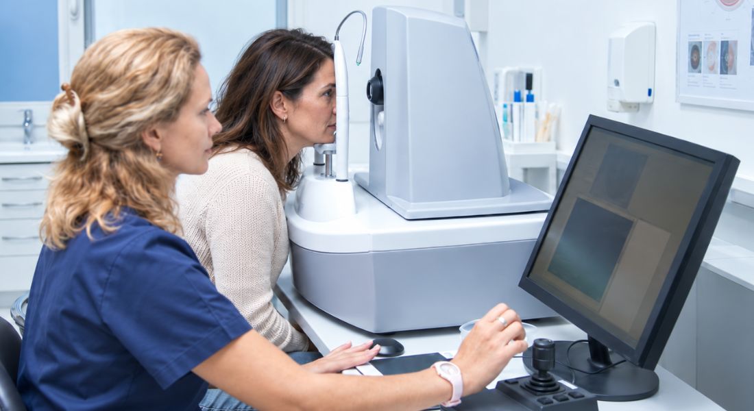
Your visual-field test checks how well you can see around the edges of your vision, not just in the centre. IIH can affect your peripheral vision even when your central vision still seems clear.
Your ophthalmologist can repeat the test to monitor changes over time. Changes in your visual fields can help show whether your vision is stable or worsening and how urgently treatment may be needed.
Clinical Tip
Your visual-field test requires concentration and can sometimes be tiring. Try to follow the instructions carefully and keep your attention on the central target throughout the test. If the result is unreliable or does not fit with your other findings, your ophthalmologist may repeat the test rather than assuming that the result represents true visual loss.
Bring your glasses or contact-lens information, current medicine list and details of previous eye or brain investigations to your appointment. Having your previous results available can help your specialist compare changes over time and assess whether your visual function is stable.
Why Are Optic Nerve Photographs Useful?
Your optic nerve photographs provide a record of how your optic discs look at each appointment. Your ophthalmologist can compare these images over time to see whether your papilloedema is improving or worsening.
Your photographs show the structure of your optic nerves, while your visual-field tests show how well they are functioning. Your doctor uses both to monitor your IIH.
Evidence Note
An elevated CSF opening pressure supports a diagnosis of IIH, but the measurement should not be interpreted on its own. UK consensus guidance uses an opening pressure above 25 cm CSF as part of the diagnostic criteria in adults, while also recognising that CSF pressure varies and that results close to the threshold can be difficult to interpret.
Your specialist therefore considers the opening pressure alongside papilloedema, visual findings, neurological examination, brain imaging, venous imaging and normal CSF composition. If the pressure result does not fit the rest of your clinical picture, your specialist may need to reassess the diagnosis rather than relying on the number alone.
What Does OCT Show in IIH?
Your OCT scan provides detailed images of the back of your eye and can measure changes around your optic nerve. Your retinal nerve fibre layer may become thicker when papilloedema is present.
Your ophthalmologist can repeat OCT scans to monitor changes over time. Your OCT results are considered alongside your visual fields and optic-disc appearance because OCT alone cannot diagnose or exclude IIH.
Research Insight
Research has shown that OCT can provide objective measurements of optic nerve swelling and can be useful for monitoring papilloedema over time. It can add structural information that complements your visual-field testing and clinical examination.
However, OCT should not be treated as a stand-alone diagnostic test for IIH. Changes on OCT need to be interpreted alongside your optic-disc examination, visual function and other investigations. Researchers continue to study how newer OCT measurements may improve the detection and monitoring of optic nerve damage.
Why Do You Need Brain Imaging Before a Lumbar Puncture?
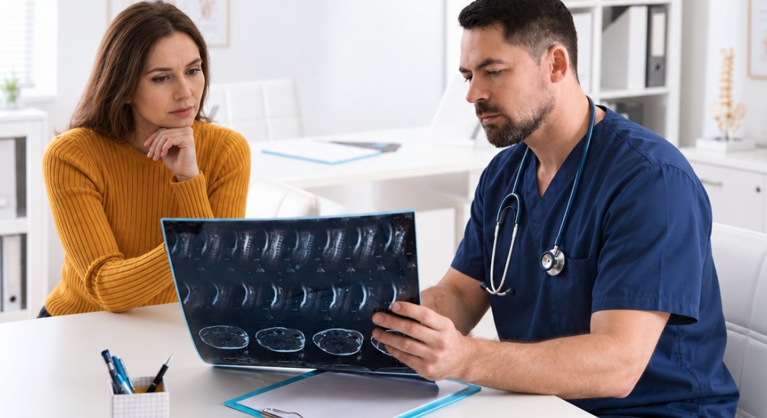
Your brain imaging helps your doctors rule out other causes of raised intracranial pressure, such as a brain mass or hydrocephalus. Your doctors also use the scan to identify conditions that could make a lumbar puncture unsafe or require a different investigation pathway.
MRI is generally preferred when available and suitable. Urgent CT may be used when MRI is not immediately available or is unsuitable, with further imaging considered when needed. Your brain and venous scans help your doctors assess the wider picture before confirming IIH.
What Can an MRI Scan Show?
Your MRI scan can show your brain, ventricles and surrounding structures in detail. Your doctors can use it to look for causes of raised intracranial pressure, such as a mass or hydrocephalus.
Your scan may also show features associated with IIH, such as changes around your optic nerves or narrowing of your venous sinuses. Your MRI findings support your assessment but cannot confirm IIH on their own.
Why Is MR or CT Venography Important?
Your MR or CT venography checks the veins that drain blood from your brain. Your doctors use it to look for problems such as cerebral venous sinus thrombosis, which can cause raised intracranial pressure, headaches and papilloedema.
Your venous imaging is important because these conditions can closely resemble IIH. Your scan helps rule out another potentially serious and treatable cause before your doctors diagnose your condition as idiopathic intracranial hypertension.
UK Guidance Note
UK consensus guidance recommends that cerebral venous imaging is included when investigating papilloedema because cerebral venous sinus thrombosis can cause raised intracranial pressure and needs to be excluded. MRI with MR venography is generally preferred when available, while CT with venography may be used when appropriate.
The same UK guidance recommends confirming papilloedema, assessing visual function and obtaining appropriate neuroimaging before diagnostic lumbar puncture. Where papilloedema is present and imaging is normal, lumbar puncture is used to measure the opening pressure and check that the CSF composition is normal.
Can a Brain Scan Confirm IIH Without a Lumbar Puncture?
Your brain scan alone usually cannot confirm IIH. Your MRI may show signs linked with raised pressure and help exclude other causes, but these findings are not specific enough on their own.
Your lumbar puncture can measure your CSF pressure and analyse the fluid when appropriate. Your doctor will consider these results alongside your symptoms, eye findings and scans.
IIH Diagnostic Tests: What Each Test Shows
| Test or assessment | What it checks | Why it matters |
| Eye examination | Your optic nerves, retina and other eye structures | Helps identify papilloedema and assess whether your vision may be at risk |
| Visual-field testing | How well you can see across your peripheral and central field of vision | Detects visual-field loss that may occur even when your central vision remains clear |
| Optic nerve photographs | The appearance of your optic discs | Provides a record that can be compared with future examinations |
| OCT scan | Detailed structural changes around your optic nerves and retina | Helps document and monitor papilloedema over time |
| MRI brain scan | Your brain and surrounding structures | Helps exclude a mass, hydrocephalus or other structural cause and may show features associated with raised pressure |
| MR or CT venography | The veins that drain blood from your brain | Helps exclude cerebral venous sinus thrombosis and assess the cerebral venous system |
| Lumbar puncture | Your CSF opening pressure and CSF composition | Provides pressure measurement and helps exclude other causes of raised intracranial pressure |
| Neurological assessment | Your cranial nerves, strength, reflexes, coordination and other neurological function | Helps identify findings that may suggest another neurological condition |
What Is a Lumbar Puncture?
Your lumbar puncture is a procedure that collects cerebrospinal fluid through a fine needle placed in your lower back. Your doctor uses a local anaesthetic to help reduce discomfort.
Your lumbar puncture can measure your CSF opening pressure and test the fluid for other conditions. Your head may ache afterwards, particularly when you are upright, but most procedures are completed safely.
How Is the Opening Pressure Measured?
Your CSF opening pressure is usually measured with a manometer while you lie on your side in the lateral decubitus position. You should be relaxed, and the reading is generally taken after the pressure level has settled. Your doctor records the opening pressure before cerebrospinal fluid is removed for testing.
Your body position, straining and anxiety can affect the reading, so your doctor considers how the measurement was taken. Your pressure can also naturally vary over time.
What Does Your Lumbar-Puncture Pressure Result Mean?

UK consensus guidance uses an opening pressure above 25 cm CSF as part of the diagnostic criteria for IIH in adults. However, readings around this threshold need to be interpreted alongside your eye findings, imaging, symptoms and the way the lumbar puncture was performed.
Your pressure reading can be affected by positioning and technique, so one number does not confirm IIH by itself. Your specialist will consider your full clinical picture.
Why Is the Cerebrospinal Fluid Analysed?
Your lumbar puncture also allows your doctors to test your cerebrospinal fluid. In typical IIH, your CSF should have a normal composition.
Your tests may check cells, protein and glucose to look for another cause of raised pressure. Your results are considered alongside your symptoms, scans and eye findings.
Myth vs Fact
| Myth | Fact |
| One test can confirm IIH. | IIH is diagnosed by bringing together your symptoms, eye findings, imaging, CSF results and exclusion of other causes. |
| A normal MRI means you cannot have IIH. | MRI may not show a structural cause while still showing features associated with raised intracranial pressure. A normal MRI does not by itself exclude IIH. |
| An MRI scan alone can diagnose IIH. | MRI can help exclude other causes and may show features associated with raised pressure, but it does not usually confirm IIH on its own. |
| OCT can diagnose IIH by itself. | OCT is useful for assessing and monitoring optic nerve swelling, but it needs to be interpreted alongside your clinical examination and visual-function testing. |
| Good central vision means IIH has not affected your eyesight. | IIH can affect your peripheral visual field while central vision remains relatively clear, so formal visual-field testing is important. |
| A raised lumbar-puncture pressure automatically proves IIH. | An elevated opening pressure supports the diagnosis, but it must be interpreted alongside your symptoms, eye findings, imaging and CSF composition. |
| A lumbar puncture is only used to measure pressure. | CSF can also be analysed for cells, protein, glucose and other findings that may help identify another cause of raised intracranial pressure. |
| Swollen-looking optic discs always mean papilloedema. | Optic discs can appear swollen because of conditions such as optic disc drusen or naturally crowded discs. Specialist assessment may be needed to distinguish papilloedema from pseudopapilloedema. |
| You only need visual testing when your vision feels blurred. | Visual-field loss can occur before you notice obvious changes in central vision, so formal visual assessment can be important even when your sight feels relatively normal. |
| Once the diagnosis is made, further eye tests are unnecessary. | Regular visual-field testing, optic-disc assessment and sometimes OCT or photographs help monitor whether your optic nerves and vision are stable. |
Key Takeaways
- IIH is diagnosed using several findings rather than one test.
- Your eye examination helps identify papilloedema and assess whether your vision is at risk.
- Formal visual-field testing is important because IIH can affect peripheral vision even when central vision remains clear.
- Optic nerve photographs and OCT can help your ophthalmologist document and monitor changes over time.
- MRI or CT brain imaging helps exclude structural causes of raised intracranial pressure.
- MR or CT venography is important for excluding cerebral venous sinus thrombosis and assessing the cerebral venous system.
- A lumbar puncture measures your CSF opening pressure and allows your CSF to be analysed.
- An elevated CSF opening pressure supports IIH but should not be interpreted in isolation.
- A normal CSF composition is part of the usual diagnostic criteria for IIH.
- Your doctors also need to consider medicines and other conditions that can cause secondary intracranial hypertension.
- If your visual symptoms are new or worsening, you should seek prompt medical or ophthalmic assessment.
- Your final diagnosis depends on how your symptoms, eye findings, imaging and CSF results fit together.
Frequently Asked Questions
- How is idiopathic intracranial hypertension (IIH) diagnosed?
Your IIH diagnosis usually involves several tests rather than one examination. Your doctors may assess your eyes, visual fields and optic nerves, use brain and venous imaging, and perform a lumbar puncture when appropriate. - Why is an eye examination important when diagnosing IIH?
Your eye examination can identify papilloedema, which is swelling of your optic nerve heads caused by raised intracranial pressure. It also helps your ophthalmologist assess whether your vision may be at risk. - Why do you need a visual-field test for IIH?
Your visual-field test checks your peripheral vision, which can be affected by IIH even when your central vision remains clear. Repeating the test can help your ophthalmologist identify whether your vision is stable or worsening. - What does OCT show in IIH?
Your OCT provides detailed images of the back of your eye and can measure changes around your optic nerve. It can help monitor papilloedema over time, but OCT alone cannot diagnose or exclude IIH. - Can an MRI scan confirm IIH?
Your MRI can help rule out other causes of raised intracranial pressure and may show features associated with IIH. However, an MRI alone cannot usually confirm IIH, so your doctor will consider it alongside your other findings. - Why might you need MR or CT venography?
Your MR or CT venography examines the veins that drain blood from your brain. It helps your doctors look for conditions such as cerebral venous sinus thrombosis, which can also cause raised intracranial pressure and papilloedema. - What is a lumbar puncture for IIH?
A lumbar puncture uses a fine needle in your lower back to collect cerebrospinal fluid and measure its opening pressure. Your doctors can also analyse the fluid to help exclude other causes of raised pressure. - What does the opening pressure result mean?
UK consensus guidance uses an opening pressure above 25 cm CSF as part of the diagnostic criteria for IIH in adults. Your specialist will still interpret the result alongside your symptoms, eye findings, imaging and how the measurement was taken. - Can your optic discs look swollen without having IIH?
Yes. Your optic discs can appear swollen for reasons other than raised intracranial pressure, including naturally crowded optic discs or optic disc drusen. Your ophthalmologist may use OCT, photographs and other tests when the diagnosis is unclear. - Why is cerebrospinal fluid analysed during a lumbar puncture?
Your cerebrospinal fluid is analysed to check for other conditions that could cause raised intracranial pressure. In typical IIH, your CSF composition should be normal, including its cells, protein and glucose.
Final Thoughts: How IIH Is Diagnosed
Diagnosing IIH involves several investigations to identify papilloedema, assess how well your optic nerves are functioning and rule out other causes of raised intracranial pressure. Your eye examination, visual fields, OCT, brain and venous imaging and, when appropriate, lumbar puncture all provide important information that needs to be considered together.
If you have headaches, pulsatile tinnitus, visual symptoms or have been told that your optic discs appear swollen, you can contact Eye Clinic London for an assessment of your vision and advice on appropriate diagnostic next steps.
References:
- Mollan, S.P., Davies, B., Silver, N.C., Shaw, S., Mallucci, C.L., Wakerley, B.R., Krishnan, A., Chavda, S.V., Ramalingam, S., Edwards, J., Hemmings, K., Williamson, M., Burdon, M.A., Hassan-Smith, G., Digre, K., Liu, G.T., Højland Jensen, R. and Sinclair, A.J. (2018) ‘Idiopathic intracranial hypertension: consensus guidelines on management’, Journal of Neurology, Neurosurgery & Psychiatry, 89(10), pp. 1088–1100. Available at: https://pmc.ncbi.nlm.nih.gov/articles/PMC6166610/
- Malhotra, K., Padungkiatsagul, T. and Moss, H.E. (2020) ‘Optical coherence tomography use in idiopathic intracranial hypertension’, Annals of Eye Science, 5, Article 7. Available at: https://pmc.ncbi.nlm.nih.gov/articles/PMC7220123/
- Lowe, M.C., Berman, G., Labella Álvarez, F., Shah, P. and Mollan, S.P. (2026) ‘How to diagnose papilloedema’, Practical Neurology, 26(4), pp. 319–330. Available at: https://pubmed.ncbi.nlm.nih.gov/41529938/
- Korsbæk, J.J., Jensen, R.H., Høgedal, L., Molander, L.D., Hagen, S.M. and Beier, D. (2023) ‘Diagnosis of idiopathic intracranial hypertension: A proposal for evidence-based diagnostic criteria’, Cephalalgia, 43(3), Article 3331024231152795. Available at: https://pubmed.ncbi.nlm.nih.gov/36786317/
- Wang, M.T.M., Bhatti, M.T. and Danesh-Meyer, H.V. (2022) ‘Idiopathic intracranial hypertension: Pathophysiology, diagnosis and management’, Journal of Clinical Neuroscience, 95, pp. 172–179. Available at: https://www.sciencedirect.com/science/article/pii/S0967586821005853