


The History of Corneal Transplantation
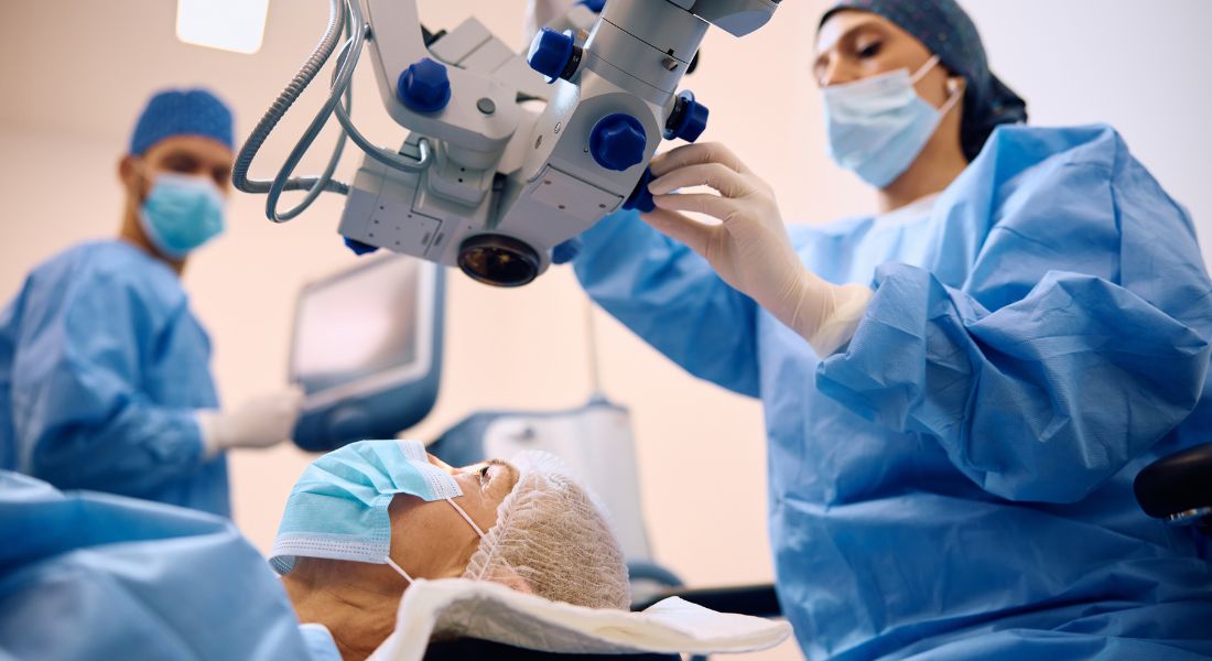
Corneal transplantation has evolved from experimental attempts into highly specialised surgery that can replace either the full thickness of your cornea or only the diseased layers. More than a century of advances in donor tissue, eye banking, microsurgery and understanding of corneal anatomy have made transplantation increasingly selective.
Today, your surgeon may consider penetrating keratoplasty, DALK, DSAEK or DMEK depending on which part of your cornea is affected. These techniques aim to restore corneal clarity while preserving as much of your healthy tissue as possible.
What Is a Corneal Transplant?
A corneal transplant, or keratoplasty, replaces damaged corneal tissue with healthy donor tissue. The aim is usually to restore clarity, improve vision or protect an eye affected by serious corneal disease.
Your surgeon may replace the entire thickness of your cornea or only the damaged layers, depending on the condition being treated. Modern techniques allow more selective transplantation while preserving healthy corneal tissue.
Early Ideas About Replacing the Cornea
The idea of restoring a damaged transparent cornea existed centuries before successful transplantation became possible. Early physicians and scientists proposed replacing or removing opaque corneal tissue, but surgical technology was not yet advanced enough.
If you had needed a corneal transplant during this early period, the available surgical techniques would not have been advanced enough to provide a reliable result. Progress in surgical instruments, anaesthesia, microscopy and understanding of the eye eventually made successful corneal transplantation possible.
Early Experimental Transplants
During the nineteenth century, surgeons attempted corneal transplantation using both human and animal tissue. Most early grafts failed because of infection, inflammation, poor surgical techniques and limited understanding of tissue healing.
If you had needed a corneal transplant during this period, these limitations would have made successful healing and long-term graft survival much more difficult. These early attempts nevertheless provided valuable experience and helped surgeons understand the challenges of replacing corneal tissue. Improvements in surgical technique and postoperative care gradually increased the chances of successful transplantation.
Eduard Zirm’s Breakthrough
On 7 December 1905, Austrian ophthalmologist Eduard Zirm performed the first successful human penetrating corneal transplant. The operation showed for the first time that transplanted human corneal tissue could remain sufficiently clear to restore useful vision.
If your cornea requires transplantation today, your operation is considerably more sophisticated than Zirm’s procedure. However, his successful graft established the clinical foundation from which modern penetrating and lamellar transplantation developed.
What Was Penetrating Keratoplasty?

Penetrating keratoplasty, or PK, replaces the full thickness of your damaged central cornea with a circular piece of donor tissue. For many decades, it became the principal technique for treating serious corneal scarring and disease.
If you need a corneal transplant, your surgeon may consider PK when the damage affects the full thickness of your cornea. The procedure can replace the damaged tissue and help restore clearer vision.
Why Did Early Transplants Often Fail?
Early surgeons did not have modern operating microscopes, fine sutures, antibiotics, corticosteroid treatment or reliable donor-tissue preservation. Their ability to recognise and manage graft rejection was also much more limited than it is today.
If you receive a transplant now, your surgeon combines refined microsurgery with carefully assessed donor tissue and structured postoperative treatment and follow-up. These developments have greatly improved the predictability of corneal graft surgery, although rejection, infection and graft failure can still occur.
The Development of Eye Banking
Reliable access to donor corneas became increasingly important as corneal transplantation expanded. Townley Paton helped establish organised eye banking in New York during the 1940s, supporting the collection, preservation and distribution of donor tissue.
- Donor access: Eye banking helps provide a more organised supply of suitable donor corneas for transplantation
- Tissue assessment: Donor corneas can be examined and assessed for suitability before they are offered for surgery
- Preservation: Specialised storage methods help maintain donor tissue until it is needed for transplantation
- Tissue distribution: Eye banks coordinate the distribution of suitable corneas to hospitals and transplant centres
- Modern transplantation: If you need a corneal transplant today, this organised system helps make suitable donor tissue more reliably available
The development of eye banking has played an important role in making corneal transplantation more organised and accessible. Today, donor corneas can be carefully assessed and preserved before surgery, helping your surgical team plan your transplant more effectively.
Better Microsurgical Techniques
Operating microscopes, finer instruments and improved sutures gave surgeons much greater control over graft positioning and wound closure. These advances helped penetrating keratoplasty become more predictable during the twentieth century.
If you need a corneal transplant, these microsurgical advances allow your surgeon to position donor tissue more precisely and close the graft securely. This has helped improve the safety and predictability of corneal transplantation.
Understanding Corneal Graft Rejection
Researchers gradually learned more about how your immune system can recognise and attack donor corneal tissue. Better recognition of graft rejection and the use of anti-inflammatory treatment improved the ability to protect transplanted corneas.
If you have a corneal transplant, recognising the warning signs of graft rejection early can help your ophthalmologist treat the problem promptly. Your follow-up care and prescribed treatment are important for protecting your transplanted cornea.
Why Replace the Full Thickness of Your Cornea?
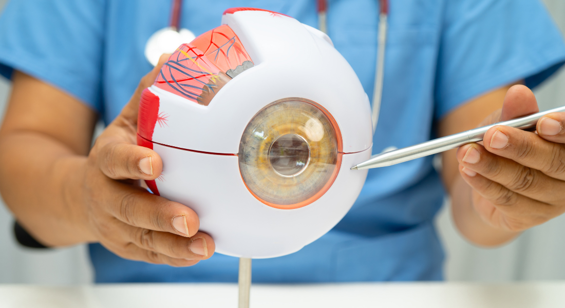
Your penetrating keratoplasty replaces the full thickness of the affected central part of your cornea. Historically, this meant that all corneal layers within the graft area were replaced even when your disease mainly affected particular layers.
Your surgeon may now be able to preserve healthy layers of your own cornea by using a lamellar transplant instead. Your most suitable procedure depends on which corneal layers are affected and the condition of the rest of your eye.
The Development of Lamellar Keratoplasty
Lamellar keratoplasty replaces only selected layers rather than the entire cornea. This more targeted approach became increasingly practical as surgical instruments and understanding of corneal anatomy improved.
If only specific layers of your cornea are damaged, your surgeon may be able to replace those layers while preserving the healthier tissue underneath. This has helped make corneal transplantation more selective and precise.
Deep Anterior Lamellar Keratoplasty
DALK removes diseased corneal stroma while preserving your own Descemet membrane and endothelial cells. Your surgeon may consider it when conditions such as keratoconus or stromal scarring affect the front and middle parts of your cornea while your endothelium remains healthy.
Because your own endothelial cells are retained, you do not face endothelial graft rejection in the same way as you would after a transplant containing donor endothelium. DALK can nevertheless have other complications, and in some operations your surgeon may need to convert to a full-thickness transplant.
The Big-Bubble Technique
In 2002, Mohammed Anwar and Klaus Teichmann described the big-bubble technique for DALK. Your surgeon injects air deep within your corneal stroma to help separate the diseased stromal tissue from the underlying Descemet membrane.
When the separation is successful, your surgeon can remove deep stromal tissue while preserving your own Descemet membrane and endothelial cells. The technique has become an important approach to DALK, although achieving the correct tissue plane remains technically demanding.
The Rise of Endothelial Keratoplasty
The development of endothelial keratoplasty changed treatment for diseases affecting the back of your cornea. In 1998, Gerrit Melles and colleagues described a posterior lamellar approach designed to replace posterior corneal tissue while leaving your anterior cornea intact.
Further developments increasingly reduced the amount of donor tissue placed inside your eye. These advances eventually led from posterior lamellar surgery to DSEK and DSAEK and then to the much thinner DMEK graft.
DSEK and DSAEK
DSEK and DSAEK are corneal transplant techniques that replace only the damaged back layers of your cornea rather than the full thickness. This can provide faster visual recovery than traditional full-thickness transplantation when the front layers of your cornea remain healthy.
Key Points About DSEK and DSAEK
| Aspect | What It Means for You | Why It Matters for You |
| Your DSEK graft | Your donor graft contains endothelium, Descemet membrane and a thin layer of posterior stroma | Your diseased endothelial tissue can be replaced without a full-thickness corneal transplant |
| Your DSAEK graft | Your donor tissue contains the same main layers but is prepared using an automated microkeratome technique | Your surgeon receives a more consistently prepared donor lenticule |
| Your partial transplant | Only the diseased back portion of your cornea is replaced | More of your healthy anterior cornea can be preserved |
| Your suitability | Your surgeon may consider this approach when your main problem is endothelial failure | You may avoid a full-thickness transplant when your front corneal layers remain suitable |
| Your recovery | Your vision may recover more quickly than after penetrating keratoplasty | Your rehabilitation can be shorter in suitable eyes |
| Your surgery | Your donor graft is positioned against the back of your cornea | Your procedure requires specialised endothelial transplant techniques |
The Development of DMEK
In 2006, Melles and colleagues described Descemet membrane endothelial keratoplasty, or DMEK. Instead of transplanting donor stroma as part of the graft, DMEK replaces only your Descemet membrane and endothelial cells.
If your corneal disease is limited to endothelial failure, this very thin graft can preserve almost all of your own corneal structure. DMEK has subsequently become an important option for conditions such as Fuchs endothelial dystrophy and selected cases of endothelial decompensation.
Why Was DMEK a Major Advance?
DMEK can provide rapid, high-quality visual rehabilitation in suitable eyes because your transplanted tissue closely matches the corneal layers that are actually diseased. Studies have also reported a relatively low risk of immune rejection compared with thicker endothelial grafts and penetrating keratoplasty.
However, your DMEK graft can partially detach after surgery. If this happens, your surgeon may need to inject another air or gas bubble to reattach it, a procedure known as rebubbling. Your individual balance of benefits and risks therefore needs to be discussed before surgery.
How Has Modern Transplantation Changed?
Modern corneal transplantation is increasingly based on replacing the part of your cornea that is diseased rather than automatically replacing its full thickness. Your surgeon may use PK when several layers are affected, DALK when your stroma is diseased but your endothelium is healthy, or DSAEK or DMEK when endothelial failure is the main problem.
This layer-specific approach can preserve more of your healthy cornea and may reduce particular complications or shorten visual rehabilitation in suitable cases. It does not mean that one procedure is best for everyone, because your diagnosis, previous surgery and other eye conditions can change which technique is safest and most appropriate.
UK Guidance Note
If you undergo corneal transplantation in the UK, your operation uses donated human corneal tissue that has been retrieved, assessed and stored through regulated tissue-banking systems. Only the corneal tissue needed for transplantation is ultimately grafted into your eye.
Your surgeon should recommend the type of transplant according to which layers of your cornea are diseased. UK centres currently perform both full-thickness and partial-thickness grafts, including DALK, DSAEK and DMEK, and your choice of procedure should be based on your diagnosis rather than simply selecting the newest technique.
Key Takeaways
- Your modern corneal transplant developed from more than a century of surgical innovation.
- Your first successful human corneal transplant was achieved by Eduard Zirm in 1905.
- Your donor tissue can now be assessed, stored and distributed through organised eye-banking systems.
- Your full-thickness transplant is known as penetrating keratoplasty.
- Your DALK procedure preserves your own Descemet membrane and endothelial cells.
- Your big-bubble DALK technique was described by Anwar and Teichmann in 2002.
- Your DSAEK procedure replaces your diseased endothelium with donor endothelium and a thin layer of supporting stroma.
- Your DMEK procedure uses an even thinner graft containing Descemet membrane and endothelial cells.
- Your DMEK recovery may be faster than after full-thickness transplantation, although graft detachment can require rebubbling.
- Your surgeon should choose the transplant according to the layers affected by your particular corneal disease.
Myth vs Fact
| Myth | What You Should Know |
| Your whole eye is replaced during a corneal transplant. | Only your diseased corneal tissue is replaced with donor corneal tissue. |
| Your modern transplant always replaces the full cornea. | Your surgeon may replace only the diseased anterior or endothelial layers. |
| Your DALK procedure uses donor endothelial cells. | Your own Descemet membrane and endothelial cells are normally preserved. |
| Your DALK cannot have any graft rejection. | Your preserved endothelium avoids endothelial graft rejection, but other graft-related immune reactions can still occur. |
| Your DSAEK and DMEK procedures are the same. | Your DSAEK graft contains supporting donor stroma, while your DMEK graft contains only Descemet membrane and endothelium. |
| Your DMEK graft always attaches after one operation. | Your graft can detach and sometimes needs an additional air or gas injection called rebubbling. |
| Your DMEK is automatically better than every other transplant. | Your most appropriate procedure depends on which parts of your cornea are diseased. |
| Your transplant permanently removes every future risk. | Your graft still requires follow-up because rejection, failure and other complications can occur. |
Frequently Asked Questions
- What is a corneal transplant?
Your corneal transplant replaces diseased or damaged corneal tissue with healthy donor tissue. Your surgeon may replace the full thickness of your cornea or only the particular layers affected by disease. - When was the first successful corneal transplant performed?
Eduard Zirm performed the first successful human penetrating corneal transplant in 1905. His operation provided an important foundation for the transplant techniques available to you today. - What is penetrating keratoplasty?
Penetrating keratoplasty replaces the full thickness of your central cornea with donor tissue. Your surgeon may consider it when disease or scarring affects several corneal layers. - Why did early corneal transplants often fail?
Your chances of successful transplantation would historically have been limited by poor tissue preservation, infection, inadequate instruments and limited understanding of graft rejection. Modern microsurgery, eye banking and postoperative treatment have addressed many of these problems. - What is lamellar keratoplasty?
Your lamellar transplant replaces selected corneal layers while preserving tissue that remains healthy. DALK and endothelial keratoplasty are important examples of this more selective approach. - What is DALK?
Your DALK procedure replaces diseased corneal stroma while retaining your own Descemet membrane and endothelial cells. It may be suitable when conditions such as keratoconus affect your corneal structure but leave your endothelium healthy. - What is DSAEK?
Your DSAEK procedure replaces diseased endothelial tissue using donor endothelium, Descemet membrane and a thin layer of supporting stroma. It is used when your endothelial cells are no longer keeping the cornea clear. - What is DMEK?
Your DMEK procedure replaces only your diseased Descemet membrane and endothelial cells with an extremely thin donor graft. It can provide rapid visual rehabilitation in suitable eyes. - Is DMEK better than a full-thickness transplant?
Not for every condition. Your DMEK may be preferable when disease is confined to the endothelium, while your surgeon may recommend DALK or penetrating keratoplasty when other corneal layers are affected. - How has corneal transplantation changed over time?
Your modern transplant can now target individual diseased layers rather than automatically replacing the whole cornea. Developments such as DALK, DSAEK and DMEK have made corneal transplantation increasingly selective.
Final Thoughts: The Evolution of Corneal Transplantation
The history of corneal transplantation shows how advances in microsurgery, donor tissue preservation and understanding of the cornea have transformed treatment. From early full-thickness transplants to modern techniques such as DALK, DSAEK and DMEK, your surgeon can now replace only the layers affected by your condition in many cases. This more targeted approach can preserve healthy corneal tissue and support improved visual recovery.
If you have corneal disease, scarring or reduced vision and are considering a corneal transplant in London, contact us at Eye Clinic London to arrange a consultation. Your cornea can be assessed in detail so your ophthalmologist can determine whether transplantation is appropriate and which treatment approach may be most suitable for you.
References
- Paton, D. (1991) ‘The founder of the first eye bank: R. Townley Paton, MD’, Refractive and Corneal Surgery, 7(2), pp. 190–194. Available at: https://pubmed.ncbi.nlm.nih.gov/2043567/
- Price, F.W. Jr and Price, M.O. (2005) ‘Descemet’s stripping with endothelial keratoplasty in 50 eyes: a refractive neutral corneal transplant’, Journal of Refractive Surgery, 21(4), pp. 339–345. Available at: https://pubmed.ncbi.nlm.nih.gov/16128330/
- Shams, M., Sharifi, A., Akbari, Z., Maghsoudlou, A. and Tajali, M.R. (2022) ‘Penetrating keratoplasty versus deep anterior lamellar keratoplasty for keratoconus: a systematic review and meta-analysis’, Journal of Ophthalmic and Vision Research, 17(1), pp. 89–107. Available at: https://pmc.ncbi.nlm.nih.gov/articles/PMC8850853/
- Singh, A., Zarei-Ghanavati, M., Avadhanam, V. and Liu, C. (2017) ‘Systematic review and meta-analysis of clinical outcomes of Descemet membrane endothelial keratoplasty versus Descemet stripping endothelial keratoplasty/Descemet stripping automated endothelial keratoplasty’, Cornea, 36(11), pp. 1437–1443. Available at: https://pubmed.ncbi.nlm.nih.gov/28834814/
- Singh, R., Gupta, N., Vanathi, M. and Tandon, R. (2019) ‘Corneal transplantation in the modern era’, Indian Journal of Medical Research, 150(1), pp. 7–22. Available at: https://pmc.ncbi.nlm.nih.gov/articles/PMC6798607/