


Can Artificial Intelligence Predict Vision Loss Before It Happens?
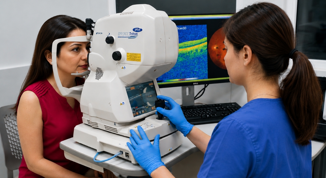
Some eye conditions can cause measurable changes to your retina, optic nerve or visual field before you notice a significant difference in your sight. Artificial intelligence is being developed to analyse these patterns and estimate whether your eye condition may have a higher risk of progressing.
AI cannot currently tell you with certainty whether you will lose vision, when this might happen or how much sight you could lose. Research involving AMD, diabetic retinopathy and glaucoma is promising, but your clinical examination, imaging and repeated follow-up remain essential when decisions are made about your care.
What Does AI Actually Predict?
AI generally estimates the probability that your eye condition will develop or worsen over a specified period. Depending on the system, your result may relate to structural progression, development of advanced disease, future visual-field changes or another defined clinical outcome.
Your prediction is not the same as knowing that you will lose sight. You should ask what outcome the model predicts, over what period and how accurately it has been tested in people similar to you.
What Information Can AI Analyse?
AI models may analyse your OCT scans, retinal photographs, visual-field tests or OCT angiography when these data are relevant to the condition being studied. Some models may also combine imaging with your age, previous results and other clinical information.
The information used varies considerably between systems. You should not assume that a model using several types of data will automatically provide a more accurate prediction for you unless it has been appropriately validated.
Why Is OCT Important for Prediction?
OCT is important because it gives your eye specialist detailed images of the retina and optic nerve, including individual layers that may not be visible during a routine examination. AI can analyse these images and detect subtle changes in fluid, drusen, retinal layers and nerve fibres.
These changes can sometimes be difficult or time-consuming to measure manually, particularly when they are small. By analysing OCT scans consistently, AI may help your specialist identify patterns that could indicate a higher risk of future disease progression.
Can AI Detect Changes Before Symptoms?
AI may identify patterns in your scans before you notice obvious changes in your sight. However, your OCT, retinal photograph or another clinical test may already show these abnormalities even without AI.
AI therefore acts mainly as an additional method of analysing your existing clinical information. Your result may help identify a pattern associated with greater progression risk, but it cannot guarantee that you will later develop symptoms or vision loss.
Predicting Advanced Macular Degeneration
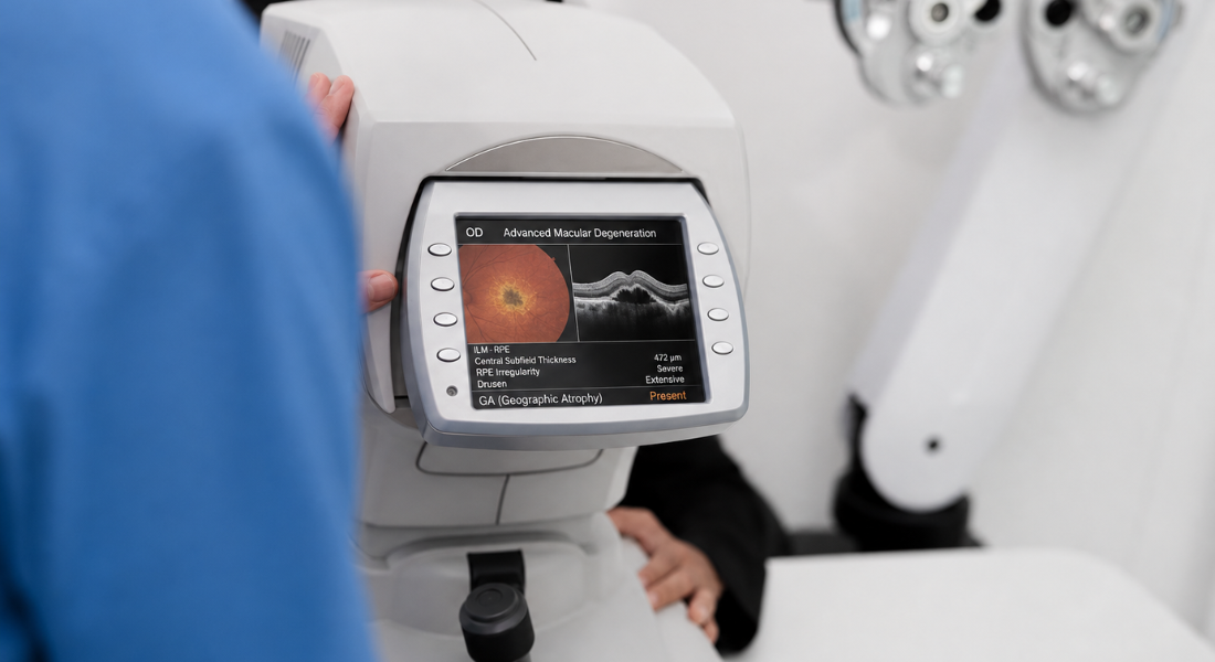
AI tools are being developed to estimate whether intermediate age-related macular degeneration may progress to advanced disease. They can analyse retinal changes that may indicate a higher risk of progression.
- Geographic atrophy: In eyes with established geographic atrophy, AI may help predict how the affected areas could enlarge over time.
- Neovascular AMD: AI tools may assess OCT patterns associated with a higher risk of future progression to neovascular AMD.
- OCT scans: These scans provide detailed images of the retina, including drusen and outer retinal changes.
- Risk assessment: AI may help your eye specialist identify whether closer monitoring or further assessment is needed.
AI can support risk assessment, but it does not replace your eye specialist’s clinical judgement. Your symptoms, scans and overall eye health are considered together.
How Accurate Are AMD Predictions?
A 2024 study evaluated a self-supervised model called Morph-SSL for predicting whether your intermediate AMD might progress to neovascular AMD using OCT scans. The model achieved an AUC of 0.779 for predicting conversion within the following six months when it was evaluated using five-fold cross-validation.
An AUC of 0.779 describes how well the model distinguished between higher-risk and lower-risk cases within the study; it does not mean that your individual prediction would be 77.9% accurate. Because the study did not establish performance through broad independent real-world validation, your AMD monitoring should continue to depend on your symptoms, examination, OCT findings and specialist assessment.
Predicting Geographic Atrophy Progression
AI can be used in research to analyse geographic atrophy and predict patterns of enlargement over time. A 2024 study showed that a deep-learning model could use a single baseline OCT scan to model future geographic atrophy lesion growth.
These models cannot currently tell you exactly how quickly your atrophy will enlarge or how your vision will change. Your ophthalmologist should continue to interpret your imaging alongside the location of the atrophy, your visual function and other clinical findings.
Diabetic Retinopathy
Diabetic retinopathy can sometimes worsen before you notice any significant change in your vision. AI can analyse retinal photographs to identify patterns in your eyes that may indicate how severe the condition is and whether you could be at greater risk of progression.
Your eye specialist can use this information alongside your usual eye examinations and other tests to understand your individual risk. This may help you receive closer monitoring when your eyes show signs that your diabetic retinopathy could worsen.
Can AI Forecast Your Diabetic Retinopathy?
A 2025 study developed DRForecastGAN, an AI system that generated synthetic future retinal images from earlier fundus photographs. Although the model showed potential for forecasting possible diabetic-retinopathy changes, it cannot predict exactly how your retina will look in the future, so regular diabetic eye screening and ophthalmology assessments remain essential.
Key Points About AI Forecasting
| Aspect | Study Details | What It Could Offer | Important Limitation |
| AI system | DRForecastGAN generated synthetic future retinal photographs | May help visualise possible disease progression | Does not show your predetermined future retina |
| Training data | 12,852 retinal images were used for training | Provided data for developing the model | AI performance depends on its training data |
| Validation | 2,734 images were used for internal validation and 8,523 for external testing | Helped assess how well the system performed | Results do not guarantee accurate individual predictions |
| Individual risk | Blood glucose, blood pressure, retinal findings and treatment can affect progression | AI may eventually support personalised monitoring | Established screening and specialist assessments remain essential |
Could AI Predict Your Sight-Threatening Diabetic Complications?
Researchers are investigating whether your retinal photographs, OCT findings and other clinical information could help estimate your risk of developing more severe diabetic eye disease. In the future, these models may provide additional information that helps your clinical team identify when you require closer assessment.
You should not currently have your screening interval or treatment changed solely because of an experimental AI prediction. Your management should continue to depend on your actual retinal findings, symptoms, established risk factors and appropriate clinical guidance.
Glaucoma
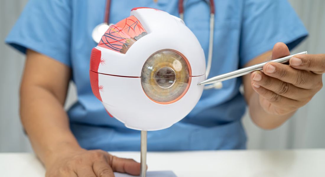
Glaucoma can gradually damage your optic nerve and peripheral vision without causing obvious symptoms in the early stages. AI is being developed to analyse your OCT scans and visual-field results to identify patterns that may indicate a higher risk of future progression.
This could help your eye specialist understand whether you may need closer monitoring or earlier intervention. However, AI cannot predict your individual outcome with certainty, so your treatment plan will still depend on your eye health, test results and clinical assessment.
What Does Current Glaucoma Research Show?
A 2026 systematic review identified 46 reports representing 43 unique studies that investigated whether AI could predict glaucoma development or progression. The models generally showed moderate to good performance when predicting outcomes such as your conversion to glaucoma, structural or functional deterioration or progression towards surgery.
However, all 43 studies were retrospective, and only seven reported testing their models on external datasets. The review also identified important limitations involving study design, generalisability, transparency and reporting of race and ethnicity. You should therefore understand that promising research performance does not mean that an AI model can yet predict your glaucoma progression reliably in routine care.
Can AI Forecast Future Visual Fields?
AI models can use your previous visual-field results, sometimes alongside OCT or other clinical information, to estimate possible future visual-field measurements. Research has demonstrated that this type of forecasting is technically possible.
Your visual-field result naturally varies between tests, and an AI-generated future field is only an estimate. You still need repeated visual-field testing so that your ophthalmologist can determine what has actually changed rather than relying on a predicted result.
Why Are Previous Changes Important?
Your previous visual-field results can show how quickly your glaucoma has been changing. A faster rate of visual-field deterioration may indicate a greater risk of subsequent progression and can help your specialist decide how closely your eyes should be monitored.
A 2025 glaucoma study found that faster visual-field deterioration during the first two years was strongly linked with later progression, supporting the value of early rates of change as a prognostic marker.
Could AI Predict Treatment Response?
Researchers are studying whether AI could estimate how your condition may respond to treatments such as retinal injections or glaucoma therapy. Your previous imaging, clinical measurements and treatment response may contain patterns that contribute to these models.
This is not yet reliable enough for AI to select your treatment independently. Your clinician should choose and adjust your treatment according to established evidence, your examination and how you actually respond over time.
Could AI Personalise Your Appointments?
AI could eventually help your specialist estimate whether you have a higher or lower risk of progression and provide additional information when planning your follow-up. If a sufficiently validated AI system identifies you as being at higher risk, its result could eventually provide additional information when your specialist considers how closely you should be monitored.
You should not have appointments delayed or reduced solely because an experimental AI model categorises you as low risk. Your follow-up interval should continue to reflect your diagnosis, previous progression, treatment, symptoms and specialist assessment.
Could Home Monitoring Improve Predictions?
Future systems may combine your clinic results with suitable home vision tests or monitoring devices. More frequent measurements could potentially give your specialist additional information about changes occurring between appointments.
A home test or AI alert should not diagnose progression independently. You should follow your existing monitoring schedule and contact your eye-care team if you notice new distortion, loss of vision or another important visual change rather than waiting for an automated alert.
What Are the Main Limitations?
Your AI prediction depends heavily on the patients, scanners, clinical settings and definitions used to develop the model. Poor-quality imaging, different equipment, unusual disease features or limited representation of people similar to you may reduce its reliability.
External validation is particularly important. A 2026 glaucoma review found that only a minority of published models had been tested on independent external datasets, showing why promising research performance cannot automatically be assumed to work equally well in your clinic.
Does AI Replace Your Eye Specialist?
No. Your AI result should support rather than replace your ophthalmologist’s judgement. Your specialist needs to consider your symptoms, examination, scans, visual-field results, medical history and previous changes before deciding what your risk means.
If an appropriately validated AI system is used during your care, you should understand what it measures and how its result affects your assessment. Your treatment should not be started, stopped or changed solely because of an experimental prediction.
Myth vs Fact
| Myth | What You Should Know |
| AI can tell you exactly whether you will become blind. | Your AI result can estimate risk but cannot predict your individual future with certainty. |
| An AI AUC of 0.779 means your prediction is 77.9% accurate. | Your AUC measures how well a model distinguishes groups and is not your personal probability of an accurate prediction. |
| AI can see retinal disease before any test can detect it. | Your OCT or retinal photograph may already show measurable changes that AI helps analyse. |
| AI-generated future retinal images show exactly what will happen to you. | Your generated image represents a modelled possibility rather than a guaranteed future appearance. |
| AI can decide how often you need glaucoma appointments. | Your follow-up should still depend on your diagnosis, measured progression and specialist assessment. |
| AI can choose the treatment that will definitely work for you. | Your response remains uncertain and your treatment must be assessed clinically. |
| A good research result means the AI will work equally well for you. | Your model may perform differently when your characteristics or clinical setting differ from its training data. |
| AI will replace your ophthalmologist. | Your specialist remains responsible for interpreting your results and making your clinical decisions. |
Key Takeaways
- Your AI result estimates risk rather than predicting your future with certainty.
- Your OCT and retinal photographs may contain measurable changes before you notice symptoms.
- Your AI system may analyse imaging, visual-field results and selected clinical information.
- Your AMD progression risk can be studied using OCT-based AI models, but these remain research tools.
- Your diabetic-retinopathy prediction may involve generated future retinal images rather than a definite forecast.
- Your glaucoma progression can sometimes be estimated using previous structural and visual-field data.
- Your previous test results remain important because your rate of change can help indicate future risk.
- Your AI result should not determine your treatment or appointment schedule independently.
- Your prediction may be less reliable if people similar to you were poorly represented in the training data.
- Your ophthalmologist remains responsible for interpreting your results and planning your care.
UK Guidance Note
If AI software provides information intended to influence your diagnosis, monitoring or treatment, it may qualify as a medical device under UK regulation depending on its intended purpose. You should not assume that an AI tool available online or incorporated into imaging software has been clinically validated to predict your individual outcome.
Your ophthalmologist should interpret any clinically used AI result alongside your established examination and testing. You should not stop medication, delay an appointment or change your treatment because of an AI prediction unless your treating clinician has advised you to do so.
FAQs
- Can AI predict whether you will lose your vision?
AI cannot currently tell you with certainty whether, when or how much vision you will lose. Your AI result may estimate the risk that a particular eye disease or clinical measurement will progress. - Which eye conditions can AI help predict?
AI prediction is being researched for conditions including AMD, diabetic retinopathy and glaucoma. The strength of evidence differs between conditions, and most progression-prediction systems remain research tools rather than routine clinical tests. - Can AI detect your eye disease before you have symptoms?
AI may identify patterns in your scans before you notice a visual change. However, your OCT, retinal photographs or other clinical tests may already reveal these abnormalities, so AI should be considered an additional analytical tool. - Which eye tests can AI analyse?
Depending on the model, your AI system may analyse OCT, retinal photographs, visual-field tests or OCT angiography. It may also combine your imaging with previous results and selected clinical information. - Can AI predict your glaucoma progression?
AI models can estimate future glaucoma progression using information such as your visual fields, OCT or clinical history. Your actual risk still needs to be assessed through repeated measurements and specialist review. - Can AI predict your macular degeneration progression?
Research models can identify OCT patterns associated with a greater likelihood of progression from intermediate to advanced AMD. Your result cannot establish with certainty whether or when that progression will occur. - Can AI predict whether your diabetic retinopathy will worsen?
Researchers have developed models that estimate progression or generate possible future retinal images. Your result remains a prediction and cannot replace your diabetic eye screening, retinal examination or established monitoring. - Could AI personalise your eye appointments?
AI may eventually provide additional risk information that helps your specialist plan follow-up. Your appointments should not currently be reduced or delayed solely because an experimental AI system considers you low risk. - What could make your AI prediction inaccurate?
Your result may be affected by poor-quality scans, different equipment, unusual disease features and under-representation of people with characteristics similar to yours. Limited external validation can also make research results difficult to apply to you. - Does AI replace your eye specialist?
No. Your ophthalmologist should consider your AI result alongside your symptoms, examination, imaging, visual fields, medical history and previous results. Your important treatment decisions should remain clinically supervised.
Final Thoughts: Understanding AI and Future Vision Loss
AI may help your eye specialist identify patterns in your scans or previous test results that are associated with a greater risk of future disease progression. Research involving your AMD, diabetic retinopathy and glaucoma is developing rapidly, but an AI prediction cannot currently tell you with certainty whether you will lose vision, when this will happen or how much your sight will change.
Your eye health should continue to be assessed using your symptoms, specialist examination, imaging, visual-field testing and previous results. If you are concerned about changes in your sight or would like your eye health assessed, you can contact Eye Clinic London to arrange an appropriate specialist consultation.
References:
- Adelpour, M. et al. (2025) ‘Short-term rates of visual field change predict glaucoma progression’, Ophthalmology Glaucoma, 8(6), pp. 560–568. Available at: https://www.sciencedirect.com/science/article/pii/S2589419625001103
- Chakravarty, A. et al. (2024) ‘Morph-SSL: Self-supervision with longitudinal morphing for forecasting AMD progression from OCT volumes’, IEEE Transactions on Medical Imaging, 43(9), pp. 3224–3239. Available at: https://pmc.ncbi.nlm.nih.gov/articles/PMC7616690/
- Qiao, H. et al. (2025) ‘Forecasting the diabetic retinopathy progression using generative adversarial networks’, Communications Medicine, 5(1), article 368. Available at: https://pmc.ncbi.nlm.nih.gov/articles/PMC12375066/
- Liang, Y.G., Fan, L., Teixeira-Pinto, A., Liew, G. and White, A.J.R. (2026) ‘A systematic review of AI for predicting glaucoma progression: challenges and recommendations towards clinical implementation’, npj Digital Medicine, 9, article 140. Available at: https://pubmed.ncbi.nlm.nih.gov/41571781/
- Mai, J., Lachinov, D., Reiter, G.S., Riedl, S., Grechenig, C., Bogunovic, H. and Schmidt-Erfurth, U. (2024) ‘Deep learning-based prediction of individual geographic atrophy progression from a single baseline OCT’, Ophthalmology Science, 4(4), article 100466. Available at: https://www.sciencedirect.com/science/article/pii/S2666914524000022