


The Latest Research on Tear Biomarkers for Diabetic Retinopathy
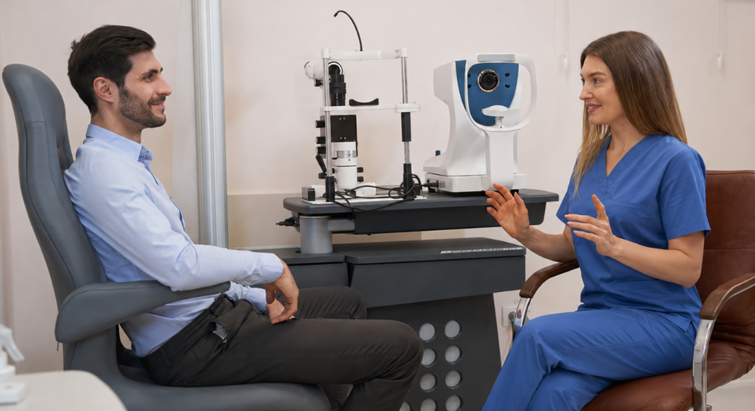
Diabetic retinopathy can develop before you notice changes in your vision. Researchers are investigating whether proteins, inflammatory molecules and genetic material in tears could provide additional information about diabetes-related eye changes.
Tear biomarkers are not currently used to diagnose or screen for diabetic retinopathy in routine care. Research remains exploratory, and retinal photography, specialist examination and other established imaging tests are still required.
Why Are Researchers Studying Tear Biomarkers?
Diabetic retinopathy can begin developing before you notice any changes in your vision. Researchers are exploring whether subtle biological signals in your tears could help detect early retinal changes or identify people who may need closer monitoring.
Tears are easy to collect and may reflect what is happening in the eye’s tissues. If useful biomarkers are identified, they could support earlier detection and help guide decisions about further testing or specialist referral.
Why Could Tears Be Useful?
Tears contain proteins, inflammatory molecules, enzymes, metabolites and genetic material. Their composition may change in association with diabetes, ocular-surface disease and diabetes-related retinal complications, making tears a useful material for exploratory biomarker research.
Tear samples can usually be collected without injections or surgery and may be suitable for repeated research measurements. However, the biological route linking retinal disease with changes in tear composition is not fully understood, and repeat testing has not yet been shown to improve routine diabetic retinopathy screening or monitoring.
Can Tear Testing Diagnose Diabetic Retinopathy Today?
Tear biomarker testing is not currently used to screen for or diagnose diabetic retinopathy in routine NHS care. In England, systematic diabetic eye screening uses digital retinal photography to look for changes at the back of the eye.
When further assessment is needed, an ophthalmology team may use a dilated retinal examination, optical coherence tomography or other retinal imaging according to the clinical findings. Tear testing should not replace or delay an invited screening appointment or specialist retinal assessment.
What Did the 2024 Proteomics Study Find?
A 2024 multicentre study analysed tear proteins in people with diabetic retinopathy, diabetes without retinopathy and healthy individuals, identifying differences that may reflect disease-related changes. The findings suggest that tear composition could provide measurable signals linked to retinal health.
| Aspect | What Was Done | Key Result | What It Means |
| Study groups | Compared three groups: diabetic retinopathy, diabetes without retinopathy and healthy participants | Enabled direct comparison across the three participant groups | Helps understand how tear proteins change with disease |
| Protein analysis | Examined 3,364 tear proteins | Large-scale proteomic assessment | Provides a detailed view of tear composition |
| Differences identified | Found 88 proteins differing between retinopathy and diabetes alone | Highlights candidate differences associated with diabetic retinopathy in this study | Suggests potential biological markers |
| Tear-based signals | Changes in protein levels linked to retinal condition | Indicates possible connection between tears and eye disease | May support future non-invasive testing |
| Clinical relevance | Explored links between tear composition and retinal changes | Offers insight into disease mechanisms | Requires further research before routine use |
Which Protein Panel Appeared Most Promising?
The researchers selected a three-protein panel consisting of SIR2, AOFB and NUD16. It distinguished diabetic retinopathy from diabetes without retinopathy with an area under the curve of 0.933 in the discovery set and 0.881 in the study’s smaller validation set.
These results show promising classification performance within this study, but they do not establish accuracy in routine screening. The panel needs external validation, standardised testing thresholds and comparison with established retinal screening before its clinical usefulness can be determined.
Why Is VEGF Important?

Vascular endothelial growth factor (VEGF) plays a key role in how your body forms new blood vessels. In diabetic retinal disease, increased VEGF can contribute to abnormal vessel growth and leakage inside your eye.
The 2025 meta-analysis found significantly higher tear VEGF concentrations in pooled groups with diabetes-related ocular complications than in participants with diabetes alone or healthy controls. However, the complication groups included conditions other than diabetic retinopathy, so the findings do not establish tear VEGF as a diabetic-retinopathy-specific marker.
What Is the Role of TNF-Alpha?
Tumour necrosis factor alpha (TNF-alpha) is involved in inflammation and can contribute to tissue damage in your body. In diabetic eye disease, higher levels of this molecule may reflect ongoing inflammatory stress affecting your eye structures.
The 2025 meta-analysis found significantly higher tear TNF-alpha concentrations in participants with diabetes-related ocular complications than in participants with diabetes alone or healthy controls. However, the analysis pooled several ocular complications, so TNF-alpha has not been shown to identify diabetic retinopathy specifically.
Are Interleukins Being Investigated?
Researchers have been studying inflammatory molecules called interleukins, including IL-6, IL-8, IL-1 beta and IL-1 receptor antagonist. These play a role in your body’s immune and inflammatory responses, which are involved in diabetic eye disease.
The 2025 meta-analysis reported higher tear IL-6 concentrations in groups with diabetes-related ocular complications. IL-8 findings were less consistent. These inflammatory markers are not specific to diabetic retinopathy and are not validated as stand-alone diagnostic tests.
What Has Research Found About Lactoferrin?
Lactoferrin helps support the protective and antimicrobial functions of your tear film. It plays a role in maintaining a stable, healthy surface environment in your eyes.
The 2025 meta-analysis found lower tear lactoferrin concentrations in the pooled ocular-complication groups. However, this finding was not specific to diabetic retinopathy, and dry eye disease, tear-film dysfunction and other ocular-surface conditions can also affect lactoferrin levels.
Which Other Tear Proteins May Be Relevant?
Researchers are exploring several additional tear proteins, including lysozyme, lipocalin-1 and markers linked to oxidative stress, metabolism, immune activity, blood-vessel formation and tissue adhesion. These molecules may reflect different biological pathways involved in diabetic eye disease.
However, most of these proteins have only been studied in one or two investigations. This means you cannot yet rely on them clinically, as they still need larger, independent studies to confirm their usefulness.
What Are Tear MicroRNAs?

MicroRNAs are small pieces of genetic material that help control how your genes are expressed. They act like regulators, influencing how cells respond to stress, inflammation and damage.
Changes in microRNA patterns in your tears may reflect processes linked to diabetic retinopathy, such as inflammation, abnormal blood-vessel activity and cellular stress. This makes them a promising area of research, although they are not yet used in routine clinical testing.
What Has Research Found About miR-15a?
A study published in December 2025 examined tear-derived exosomal miR-15a in 135 participants, including 36 with diabetic retinopathy, 50 with diabetes without retinopathy and 49 healthy controls. MiR-15a expression differed between participants with diabetes and healthy controls.
However, the study did not find a statistically significant difference between the diabetic retinopathy and diabetes-without-retinopathy groups. The result suggests that tear miR-15a may reflect diabetes-related biological changes, but it has not been shown to identify whether a person with diabetes has diabetic retinopathy.
What Has Research Published in 2026 Added?
A 2026 expert perspective concluded that tear-fluid biomarkers still face important barriers before they can enter routine clinical practice. These include inconsistent collection and analysis methods, a lack of validated reference ranges and diagnostic thresholds, limited independent clinical validation and uncertainty about how tests would fit into existing care pathways.
The authors emphasised that future research must move beyond biomarker discovery towards standardisation, reproducibility and testing in real-world clinical populations. This is particularly important for diabetic retinopathy, where any proposed tear test would need to be compared directly with established retinal screening and assessment.
Why Are Exosomes Important?
Exosomes are tiny particles released by cells that carry proteins, lipids and genetic material such as microRNAs. They act as messengers, allowing cells to communicate with each other.
Exosomes can protect some of their molecular contents from degradation, which makes exosomal proteins and microRNAs attractive candidates for biomarker research. However, this potential stability does not by itself establish that exosomal tear markers are diagnostically accurate, reproducible or specific to diabetic retinopathy.
Why Are Multi-Biomarker Panels Favoured?
Diabetic retinopathy involves several overlapping biological processes, including inflammation, vascular leakage, oxidative stress and neural injury. No individual tear marker has yet been shown to capture these processes reliably enough for routine diagnosis.
Researchers therefore investigate panels that combine several proteins or genetic markers. Combining signals may improve classification within a study, as seen with the three-protein panel reported in 2024. However, more complex panels can also overfit small datasets and must be tested independently in larger, representative clinical populations.
How Could Artificial Intelligence Help?

Artificial intelligence can help analyse complex tear biomarker patterns by assessing multiple signals at the same time. It can identify subtle relationships between proteins, genetic material and inflammatory markers that may be difficult to interpret manually.
- Pattern recognition: AI can detect complex relationships between multiple biomarkers that may not be obvious through standard analysis
- Early detection: Analysing combined signals may help identify potential disease patterns or risks at an earlier stage
- Data integration: AI can process large amounts of biological data to provide more comprehensive insights
- Decision support: These systems may assist clinicians in interpreting results and planning care more effectively
- Ongoing development: AI tools require large datasets and careful validation before being used in routine clinical practice
Machine-learning methods may eventually help researchers interpret complex tear-biomarker panels. However, no AI model based on tear biomarkers is currently validated for routine diabetic-retinopathy screening, diagnosis or treatment decisions. Models require independent testing in large and diverse populations, transparent performance reporting and comparison with established retinal assessment.
Could Tear Tests Improve Early Detection?
Researchers hope that tear tests could eventually help identify people with diabetes who need retinal imaging or closer monitoring. Such a test would need to perform reliably across different diabetes types, ethnic groups, retinopathy stages and ocular-surface conditions before it could be considered for population screening.
Current studies have not shown that tear testing detects diabetic retinopathy earlier or more reliably than established retinal screening. Prospective studies must follow people over time and demonstrate that using the test improves referral decisions or patient outcomes.
What Are the Main Research Limitations?
Studies differ in how tears are collected, stored and analysed and in how diabetes-related eye complications are defined. Dry eye disease, ocular-surface inflammation, eye drops, systemic inflammation and reflex tearing may also alter biomarker measurements and make results difficult to compare.
The 2025 meta-analysis included 19 studies and 1,413 participants but found substantial heterogeneity and evidence of publication bias. It also grouped several diabetes-related ocular complications together, meaning that findings for VEGF, TNF-alpha and other markers should not automatically be treated as specific to diabetic retinopathy.
Myth vs Fact
| Myth | Fact |
| Tear testing can currently diagnose diabetic retinopathy. | Tear biomarkers remain research tools and are not part of routine diagnosis. |
| A raised tear VEGF result confirms retinal disease. | VEGF is non-specific and can be influenced by several ocular processes. |
| Tear changes always come directly from the retina. | The biological route between retinal disease and tear composition is not fully established. |
| A promising AUC means a test is ready for patients. | Performance must be confirmed independently in larger clinical populations. |
| MiR-15a clearly distinguishes diabetic retinopathy from diabetes alone. | The 2025 study did not find a significant difference between those two groups. |
| Artificial intelligence removes the need for clinical assessment. | Machine learning can analyse patterns but still requires clinical validation and oversight. |
| Tear testing could replace retinal photographs. | Retinal photography remains the established NHS screening method. |
Key Takeaways
- Tear biomarkers for diabetic retinopathy remain at the research stage.
- Retinal photography remains the established NHS screening method.
- The biological connection between retinal disease and tear changes is not fully understood.
- A 2024 study identified a promising three-protein tear panel, but it involved a relatively small population.
- A 2025 meta-analysis identified VEGF and TNF-alpha as promising markers of diabetic ocular complications, not diabetic retinopathy alone.
- Tear miR-15a may reflect diabetes-related changes but did not clearly distinguish retinopathy from diabetes without retinopathy.
- Collection methods, dry eye disease and ocular surface inflammation can affect results.
- Larger prospective studies are needed before tear testing can enter routine care.
Frequently Asked Questions
- What Are Tear Biomarkers in Diabetic Retinopathy?
Tear biomarkers include proteins, inflammatory molecules, metabolites and genetic material that may change in people with diabetes-related eye disease. Research has not yet established a tear marker that can independently diagnose diabetic retinopathy. - Why Are Researchers Interested in Tear Testing?
Your tears are easy to collect and may reflect what is happening inside your eye without invasive procedures. This makes them a promising option for repeated monitoring and early screening research. - Can Tear Biomarkers Diagnose Diabetic Retinopathy Now?
Tear biomarker testing is not part of routine diabetic retinopathy screening or diagnosis. Digital retinal photography and specialist retinal assessment remain essential. - What Did Recent Research on Tear Proteins Find?
A 2024 study found differences in 88 tear proteins between people with diabetic retinopathy and people with diabetes without retinopathy. A three-protein panel performed well within the study, but larger independent validation is needed. - Which Tear Biomarkers Are Most Promising?
VEGF, TNF-alpha, IL-6 and several protein panels have shown potential. However, many are non-specific, and studies have used different populations and collection methods. - What Role Do Inflammatory Markers Play in Tear Testing?
Inflammatory markers such as interleukins and TNF-alpha may reflect inflammatory activity associated with diabetes-related ocular complications. However, they are not specific to retinal damage and cannot currently determine whether someone has diabetic retinopathy. - What Are Tear MicroRNAs and Why Do They Matter?
MicroRNAs regulate gene expression and can be carried within extracellular vesicles in tears. A 2025 miR-15a study found diabetes-related changes but did not clearly distinguish diabetic retinopathy from diabetes without retinopathy. - How Could Artificial Intelligence Support Tear Biomarker Research?
Machine-learning methods can identify patterns across several biomarkers and helped select the three-protein panel reported in a 2024 study. These models still need external clinical validation before they can support patient care. - Could Tear Testing Help Detect Diabetic Retinopathy Earlier?
It is not yet known whether tear testing can detect diabetic retinopathy earlier than retinal photography. Prospective studies following patients over time are needed to answer this question. - What Are the Limitations of Tear Biomarker Research?
Most studies are small, cross-sectional and methodologically varied. Tear collection, dry eye disease, ocular inflammation and laboratory processing may all affect the results.
Final Thoughts: The Future of Tear Biomarkers
Research into tear biomarkers is opening new possibilities for how diabetic retinopathy could be detected and monitored in the future. By analysing proteins, inflammatory markers and genetic material in your tears, scientists are exploring non-invasive ways to identify early retinal changes. Although biomarkers such as VEGF, TNF-alpha and microRNAs show promise, further large-scale studies are needed before they can be used reliably in clinical practice.
Digital retinal photography remains the established NHS screening method, while clinical retinal examination, optical coherence tomography and other imaging may be used for further assessment and monitoring when indicated.
If you have ongoing eye discomfort or would like to learn more about tear biomarkers in London, you can contact us at the Eye Clinic London to arrange a comprehensive assessment.
References
- Fan, Z., Hu, Y., Chen, L., Lu, X., Zheng, L., Ma, D. et al. (2024) ‘Multiplatform tear proteomic profiling reveals novel non-invasive biomarkers for diabetic retinopathy’, Eye, 38(8), pp. 1509–1517. Available at: https://pubmed.ncbi.nlm.nih.gov/38336992/
- Gijs, M., Enríquez-de-Salamanca, A., Hagan, S., Grzybowski, A. and Versura, P. (2026) ‘Recent developments in tear fluid biomarkers: an expert perspective on clinical translation’, Current Eye Research, published online 16 May 2026, pp. 1–9. Available at: https://pubmed.ncbi.nlm.nih.gov/42141967/
- Mahmud, N.M., Jie, L.Y., Singh, S., Ahmad, M.H.Z., Khaliddin, N., Mohamad, N.F. et al. (2025) ‘Tear-derived exosomal miR-15a as new diagnostic tool for diabetic retinopathy’, Journal of Visualized Experiments, 226, article 66687. Available at: https://pubmed.ncbi.nlm.nih.gov/41554023/
- NHS England (2025) Diabetic eye screening: guidance on camera approval. Published 21 May 2025. Available at: https://www.gov.uk/government/publications/diabetic-eye-screening-approved-cameras-and-settings/diabetic-eye-screening-guidance-on-camera-approval
- NHS England (2025) Optical coherence tomography (OCT) in diabetic eye screening surveillance clinics. Published 16 April 2025. Available at: https://www.gov.uk/government/publications/diabetic-eye-screening-optical-coherence-tomography-in-surveillance/optical-coherence-tomography-oct-in-diabetic-eye-screening-des-surveillance-clinics-starting-1-october