


Can Your Eyes Reveal Early Signs of Alzheimer’s Disease?

Biological changes associated with Alzheimer’s disease may begin years before noticeable memory or thinking difficulties develop. Researchers are investigating whether structural, vascular or biochemical changes in the retina could provide additional clues about these early processes.
Studies using OCT, OCT angiography, retinal photography and experimental light-based imaging have reported promising differences between research groups. However, retinal imaging cannot currently diagnose, predict or exclude Alzheimer’s disease and should not replace assessment by a memory specialist.
Why Are Researchers Studying the Eyes?
The retina is part of the central nervous system and contains nerve cells, supporting cells and small blood vessels. Researchers are investigating whether changes associated with neurodegeneration or vascular dysfunction in the brain may also be reflected in retinal structure or circulation.
The relationship is not direct enough to describe the eye as a diagnostic window into the brain. Retinal findings can be affected by many eye and general health conditions, so any possible Alzheimer’s-related pattern requires careful validation.
Can Alzheimer’s Begin Before Symptoms?
Amyloid and tau changes associated with Alzheimer’s disease may begin many years before a person develops noticeable cognitive symptoms. This period is often described as preclinical Alzheimer’s disease in research settings.
An abnormal biomarker does not show exactly when symptoms will develop, and some people with biological changes remain cognitively unimpaired for many years. Testing people without symptoms is not currently recommended through routine retinal imaging.
Can an Eye Test Diagnose Alzheimer’s Today?
No retinal scan, photograph or routine eye examination can currently diagnose, predict or rule out Alzheimer’s disease. Research studies have identified group-level associations, but no retinal threshold or imaging model has been validated for routine clinical diagnosis.
An eye examination may identify eye disease that affects vision, but concerns about memory, thinking or behaviour should be discussed with a GP or memory service rather than assessed through an eye scan alone.
What Is Optical Coherence Tomography?
Optical coherence tomography, or OCT, creates detailed cross-sectional images of your retinal layers. It allows clinicians to examine the structure of your retina with high precision.
Researchers use OCT to measure nerve-fibre thickness, macular structure and other subtle tissue changes. Researchers are investigating whether combinations of these measurements are associated with Alzheimer’s disease or cognitive impairment, but they cannot currently identify the cause of retinal changes in an individual patient.
Does Alzheimer’s Cause Retinal Thinning?
Some studies have reported differences in the retinal nerve fibre layer, ganglion-cell complex and other inner retinal structures among people with cognitive impairment or Alzheimer’s dementia. However, the affected layers and size of the differences vary substantially between studies.
Retinal thinning is not specific to Alzheimer’s disease and may also occur with normal ageing, glaucoma, short-sightedness, optic nerve disease and other neurological conditions. It cannot currently be used as an independent diagnostic sign.
What Did a 2025 OCT Study Find?
A 2025 study explored whether advanced image analysis of OCT scans could better distinguish between cognitive conditions than traditional measurements. The findings suggest that newer analytical features may improve how researchers identify patterns linked to Alzheimer’s disease, although this approach is not yet used in routine clinical diagnosis.
Key Findings From the Study
| Aspect | What Was Done | Key Result | What It Means |
| Study population | 82 participants: cognitively normal, mild cognitive impairment and Alzheimer’s dementia | Included a range of cognitive states | Indicates stronger classification performance than retinal thickness alone |
| OCT analysis method | Used experimental spatial-correlation features instead of just retinal thickness | Analysed patterns within OCT images | Looked beyond standard measurements |
| Alzheimer’s dementia classification | AUC of 0.935 using new features | Higher than 0.795 with thickness alone | Suggests improved discrimination in this group |
| Mild cognitive impairment classification | AUC of 0.830 using new features | Higher than 0.705 with thickness alone | Indicates better identification than traditional methods |
| Comparison with standard approach | Compared new analysis with conventional thickness measurements | New method showed stronger performance | Highlights potential added value of advanced analysis |
| Clinical relevance | Results apply to the study population only | Not yet a diagnostic test used in practice | Requires further research and validation |
What Is OCT Angiography?
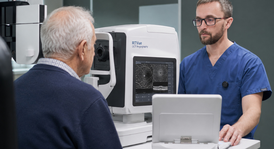
OCT angiography, or OCTA, creates images of blood flow within the small vessels of the retina without requiring an injected dye. Researchers use measurements such as vessel density, capillary patterns and the size of the foveal avascular zone when comparing groups with and without cognitive impairment.
These measurements are not specific to Alzheimer’s disease and can be affected by ageing, diabetes, high blood pressure, glaucoma, image quality and other factors. OCTA cannot currently assess your cognitive health or determine whether you have Alzheimer’s disease.
What Vascular Changes Have Been Found?
A 2025 cross-sectional study analysed one eye from each of 357 participants: 44 in a dementia group, 139 with mild cognitive impairment and 174 healthy controls. It reported reductions in vessel density around the fovea and differences in some inner retinal measurements.
The groups differed in age, sex and cognitive scores, and many participants had coexisting eye conditions. The findings show an association rather than proving that vascular changes were caused specifically by Alzheimer’s disease or could predict its development.
How Is Artificial Intelligence Being Used?
Artificial intelligence can analyse combinations of structural and vascular features within retinal images. In research studies, these systems are trained to distinguish between predefined groups, such as people with diagnosed Alzheimer’s disease, mild cognitive impairment and healthy controls.
A model’s performance within a study does not show that it can diagnose an individual patient. Before clinical use, models require independent testing in representative memory-clinic populations and validation across different scanners, imaging protocols and health conditions.
What Did the Eye-AD Study Show?
The 2024 Eye-AD study used 5,751 OCT angiography images from 1,671 participants. Its AI model reported an external AUC of 0.9007 when distinguishing early-onset Alzheimer’s disease from controls and 0.8037 when distinguishing mild cognitive impairment.
The model classified groups that had already been defined for research; it did not show that OCTA can predict which symptom-free people will later develop Alzheimer’s disease. Performance may also change when the model is used with different populations, scanners and diagnostic pathways.
Can Ordinary Retinal Photographs Be Used?
A 2022 multicentre study developed an AI model using colour retinal photographs from people with Alzheimer’s dementia and controls. Its internal performance was high, while AUC values across separate test datasets ranged from approximately 0.73 to 0.91.
The variation between datasets demonstrates the challenge of applying an algorithm to new populations. The study classified established Alzheimer’s dementia rather than detecting preclinical disease years before symptoms.
What Is Hyperspectral Retinal Imaging?
Hyperspectral retinal imaging measures how different wavelengths of light are reflected by your retinal tissue. It captures detailed spectral patterns that may reveal subtle biochemical changes.
Researchers are studying whether these patterns in your retina are linked to amyloid beta or other Alzheimer’s-related processes. This approach may offer another non-invasive way to investigate early disease changes.
How Accurate Is Hyperspectral Imaging?
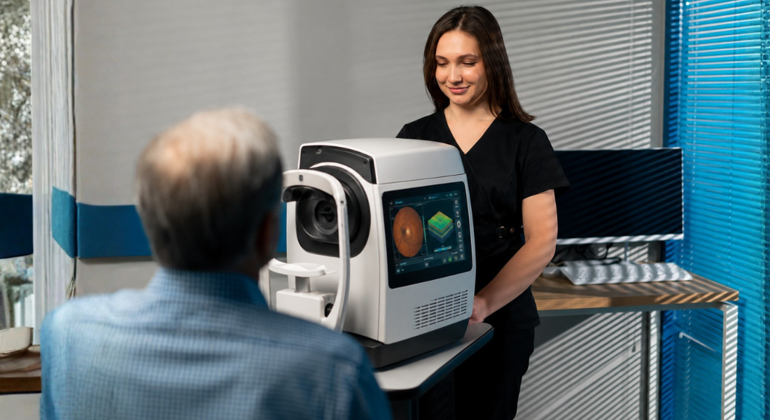
A 2025 memory-clinic study assessed whether hyperspectral retinal imaging could distinguish 57 people classified as amyloid-positive or amyloid-negative using cerebrospinal-fluid biomarkers. Its best-performing approach reported a mean AUC of 0.77, sensitivity of 0.73 and specificity of 0.55.
The relatively low specificity means that many people without amyloid could receive a positive result. Hyperspectral imaging therefore remains experimental and cannot currently confirm or exclude Alzheimer’s pathology.
Can Amyloid Be Seen in the Retina?
Researchers have used fluorescent compounds and specialised cameras to investigate possible amyloid-related deposits in the retina. The identity, origin and clinical meaning of these signals remain under investigation.
A 2025 retrospective study analysed 26 adults with and without cognitive impairment. It found associations between greater perivascular retinal amyloid measurements and poorer performance in some memory domains, but the small and demographically limited study did not validate a diagnostic retinal amyloid test.
What Did 2026 Research Show?
Erectile difficulties are a recognised side effect of treatments such as prostate surgery, radiotherapy and hormone therapy. These changes do not mean your husband is no longer attracted to you or interested in your relationship.
- Treatment effects: Erectile difficulties can occur after prostate cancer treatments and are often related to physical changes rather than a lack of attraction
- Emotional impact: Your husband may feel embarrassed, frustrated or worried about how these changes affect your relationship
- Physical affection: Remembering that intimacy does not always have to lead to sex can help reduce pressure for both of you
- Open communication: Talking honestly about concerns and feelings can help you stay connected during recovery
- Professional support: Your healthcare team can discuss available treatments and support options for sexual difficulties
Erectile difficulties can be challenging for both partners, but they do not define the strength of your relationship. With patience, reassurance and open conversations, you can continue to maintain closeness while adjusting to changes in intimacy.
Can Retinal Changes Reveal Mild Cognitive Impairment?
Some studies have found subtle retinal structural or vascular differences among groups with mild cognitive impairment. However, mild cognitive impairment is a clinical syndrome with several possible causes and does not always progress to Alzheimer’s dementia.
The retinal differences are generally smaller and less consistent than those reported in established dementia. A retinal scan cannot currently determine whether mild cognitive impairment is caused by Alzheimer’s disease.
Are These Retinal Changes Specific to Alzheimer’s?
No. Retinal thinning, altered vessel density and changes in the foveal avascular zone may also be associated with ageing, diabetes, high blood pressure, cardiovascular disease, glaucoma, macular disease and other neurological conditions.
Researchers must therefore account for eye health, medication, vascular risk, image quality and scanner type. Without this context, an apparent Alzheimer’s-related signal may reflect another condition.
Why Do Research Results Differ?
Studies differ in their imaging devices, scanning areas, image-quality standards, processing methods and definitions of Alzheimer’s disease or mild cognitive impairment. Some use biomarker-confirmed disease, while others rely mainly on clinical diagnoses or health records.
Many studies are small or cross-sectional and compare clearly diagnosed patients with healthy controls. This can produce better performance than would be expected among people attending a real memory clinic with overlapping symptoms and medical conditions.
What Tests Currently Support Alzheimer’s Diagnosis?

In UK practice, dementia assessment may include a detailed history, information from someone who knows you well, cognitive and neurological assessment, medication review, blood tests and structural brain imaging. NICE recommends offering structural imaging to look for other possible causes and help identify the dementia subtype unless dementia is already well established and the subtype is clear.
If Alzheimer’s disease is suspected but the diagnosis remains uncertain, NICE recommends considering FDG-PET, perfusion SPECT if FDG-PET is unavailable, or examination of cerebrospinal fluid for amyloid and tau biomarkers when knowing the subtype would affect management. Blood biomarkers such as phosphorylated tau are being evaluated for wider NHS use, while retinal imaging is not an established Alzheimer’s diagnostic biomarker in current UK guidance.
The 2024 Alzheimer’s Association criteria describe Alzheimer’s disease biologically and recognise sufficiently accurate amyloid PET, cerebrospinal-fluid and blood-based tests within their biomarker framework. However, these international diagnostic and staging criteria are not a UK clinical-practice guideline and should not be taken to mean that every test is routinely available or suitable as a standalone NHS assessment.
Myth vs Fact
| Myth | Fact |
| An OCT scan can diagnose Alzheimer’s disease. | OCT measures retinal structures but cannot identify the cause of any difference. |
| Retinal thinning is specific to Alzheimer’s. | Ageing, glaucoma, short-sightedness and other conditions can also affect retinal thickness. |
| A high study AUC means the test is ready for patients. | A model must be independently validated in representative clinical populations. |
| AI can see Alzheimer’s disease directly in the retina. | AI identifies statistical image patterns and does not directly observe brain pathology. |
| Mild cognitive impairment always becomes Alzheimer’s dementia. | It has several causes, and some people remain stable or improve. |
| Amyloid can already be diagnosed through an eye test. | Retinal amyloid imaging remains experimental. |
| A normal retinal scan rules out Alzheimer’s disease. | No current retinal scan can exclude Alzheimer’s disease. |
| Eye clinics can screen symptom-free people for Alzheimer’s. | Retinal screening for Alzheimer’s is not part of routine UK clinical guidance. |
Key Takeaways
- Retinal imaging for Alzheimer’s disease remains a research field.
- OCT can measure retinal layers but cannot determine the cause of thinning.
- OCT angiography can assess retinal blood vessels without injecting dye.
- Retinal changes are also influenced by ageing, diabetes, glaucoma and vascular disease.
- High AUC results from selected studies do not prove that a test is ready for screening.
- AI models must be tested in independent and clinically representative populations.
- Retinal amyloid and hyperspectral imaging remain experimental.
- No routine eye scan can currently diagnose or rule out Alzheimer’s disease.
- UK diagnosis still depends on clinical assessment and established specialist investigations.
Frequently Asked Questions
- Can an eye test diagnose Alzheimer’s disease?
No. Retinal scans and photographs are being studied, but none can currently diagnose or rule out Alzheimer’s disease. Diagnosis requires an appropriate clinical and specialist assessment. - Why are researchers studying the retina?
The retina contains nerve cells and small blood vessels and is part of the central nervous system. Researchers are investigating whether changes in retinal structure or circulation are associated with processes occurring in the brain. - What is OCT?
Optical coherence tomography creates detailed cross-sectional images of the retina. It can measure retinal layers but cannot determine whether a difference is caused by Alzheimer’s disease. - What is OCT angiography?
OCT angiography maps blood flow in the small retinal vessels without injecting dye. Researchers are studying vessel density and other measurements, but they are not established Alzheimer’s diagnostic markers. - Does Alzheimer’s disease cause retinal thinning?
Some studies have reported retinal thinning in groups with Alzheimer’s disease or cognitive impairment. The findings are inconsistent and are not specific enough to diagnose an individual patient. - Can artificial intelligence detect Alzheimer’s from an eye scan?
AI models have distinguished research groups with promising AUC values. They still require further independent testing and cannot currently be used as standalone clinical diagnostic tools. - Can amyloid be detected in the retina?
Experimental imaging has identified signals described as possible retinal amyloid. Their biological meaning and diagnostic reliability have not yet been established. - Can retinal imaging detect Alzheimer’s before symptoms?
This has not been proven. Most studies classify people who already have cognitive impairment or diagnosed dementia rather than following symptom-free people to determine who later develops Alzheimer’s disease. - Are retinal changes specific to Alzheimer’s disease?
No. Ageing, glaucoma, diabetes, high blood pressure, macular disease and other neurological or vascular conditions can produce similar findings. - What should you do if you are worried about your memory?
Contact your GP if you or someone close to you has noticed persistent changes in memory, thinking, communication or everyday functioning. An eye examination cannot replace a memory assessment.
Final Thoughts: Retinal Imaging and Alzheimer’s Research
Retinal imaging is a promising research area because it allows scientists to study nerve layers, blood vessels and light-based tissue signals non-invasively. Studies using OCT, OCT angiography, retinal photography, hyperspectral imaging and artificial intelligence have reported differences between research groups, but the findings remain inconsistent and are not specific enough for diagnosis or screening.
No routine eye scan can currently diagnose, predict or rule out Alzheimer’s disease. Concerns about persistent changes in memory, thinking, communication or everyday functioning should be discussed with a GP or memory service. If you would like to discuss your eye health concerns with an experienced specialist, get in touch with us at the Eye Clinic London today.
References
- Dallora, A.L., Alexander, J., Palesetti, P.P., Guenot, D., Selvander, M., Berglund, J.S. and Behrens, A. (2025) ‘Hyperspectral retinal imaging to detect Alzheimer’s disease in a memory clinic setting’, Alzheimer’s Research & Therapy, 17, article 232. Available at: https://pubmed.ncbi.nlm.nih.gov/41153055/
- Jin, Z., Wang, X., Lang, Y., Song, Y., Zhan, H., Shama, W. et al. (2025) ‘Retinal optical coherence tomography intensity spatial correlation features as new biomarkers for confirmed Alzheimer’s disease’, Alzheimer’s Research & Therapy, 17, article 33. Available at: https://pubmed.ncbi.nlm.nih.gov/39893456/
- Hao, J., Kwapong, W.R., Shen, T., Fu, H., Xu, Y., Lu, Q. et al. (2024) ‘Early detection of dementia through retinal imaging and trustworthy AI’, npj Digital Medicine, 7, article 294. Available at: https://pmc.ncbi.nlm.nih.gov/articles/PMC11491446/
- Ibrahim, Y., Macerollo, A., Sardone, R., Shen, Y., Romano, V. and Zheng, Y. (2025) ‘Retinal microvascular density and inner thickness in Alzheimer’s disease and mild cognitive impairment’, Frontiers in Aging Neuroscience, 17, article 1477008. Available at: https://pmc.ncbi.nlm.nih.gov/articles/PMC11906703/
- Ge, Y.-J., Xu, W., Ou, Y.-N., Qu, Y., Ma, Y.-H., Huang, Y.-Y. et al. (2021) ‘Retinal biomarkers in Alzheimer’s disease and mild cognitive impairment: a systematic review and meta-analysis’, Ageing Research Reviews, 69, article 101361. Available at: https://www.sciencedirect.com/science/article/pii/S1568163721001082