

Keratoconus in Asian Patients: Are There Differences in Presentation?
Keratoconus is a condition where the cornea gradually becomes thinner, weaker, and more cone-shaped over time. You may find that because the cornea plays an important role in focusing light into the eye, these structural changes can significantly affect visual clarity and overall quality of vision.
As the cornea changes shape, you might notice symptoms such as blurred vision, ghosting of images, glare, haloes around lights, and increasing astigmatism. These symptoms can make everyday tasks like reading, driving, or using screens more difficult, especially in low-light conditions.
Research suggests that keratoconus may be more commonly reported in some Asian populations compared with some white European populations. You might also find that studies describe differences in age at presentation, risk factors, and clinical patterns, although these findings can vary depending on the study design and population examined.
This does not mean that every Asian patient is at high risk. You may find it more helpful to focus on individual symptoms such as frequent prescription changes, distorted vision, strong astigmatism, eye rubbing, allergies, or a family history of keratoconus, all of which should be assessed properly by an eye care professional.
What Is Keratoconus?
Keratoconus affects the cornea, which is the clear front “window” of the eye. You may find that in a healthy eye, the cornea has a smooth, dome-like shape that helps focus light clearly onto the back of the eye.
In keratoconus, the cornea gradually becomes thinner and begins to bulge forward into a cone-like shape. You might notice that this change in shape affects how light enters the eye, leading to blurred, distorted, or irregular vision that can be difficult to fully correct with standard glasses.
The condition often begins in the teenage years or early adulthood, although it can sometimes be diagnosed later. You may find that early detection is important because the condition can progress over time, and regular eye checks can help monitor any changes in the cornea.
Why Asian Patients Are Discussed in Keratoconus Research
Asian patients are often discussed in keratoconus research because several studies have reported higher rates in some Asian populations compared with certain white populations. You may find that this has led clinicians and researchers to pay closer attention to patterns of risk, early signs, and diagnosis in these groups.
A UK study reported keratoconus prevalence of 229 per 100,000 in Asian patients aged 10–44 years, compared with 57 per 100,000 in white patients. You might notice that this shows a clear difference in reported prevalence within that specific study population, although results can still vary depending on methods and setting.
Another UK study found a relative incidence of keratoconus of 9.22 to 1 in Asian patients compared with white patients. You may find that findings like this highlight the importance of careful screening and awareness, particularly in younger patients who show rapidly changing prescriptions or increasing astigmatism.
These findings suggest that clinicians should remain alert to possible keratoconus in Asian patients, especially when early symptoms are present. You might notice that the emphasis is on early detection and monitoring rather than assuming risk based on ethnicity alone.
What the Research Says About Prevalence
Keratoconus prevalence can vary widely between different Asian regions. You may find that a global review has reported differences across sub regions, with generally lower estimates in East Asia and higher estimates in West Asia and South Asia.
This highlights that it is not accurate to treat all Asian patients as one single risk group. You might notice that South Asian, East Asian, Southeast Asian, West Asian, and Middle Eastern populations can each show different reported prevalence rates due to a mix of genetic, environmental, and healthcare-related factors.
The practical message is that while background can help guide clinical awareness, diagnosis should always be based on individual assessment. You may find that corneal scans and eye examinations provide the most reliable way to understand personal risk, rather than relying on broad population categories.
Why Prevalence Numbers Differ Between Studies
Keratoconus prevalence numbers can vary significantly because different studies use different definitions, diagnostic tools, and population groups. You may find that older research often relied on symptoms or basic clinical examination, which could miss early or mild cases.
In contrast, newer studies frequently use advanced corneal imaging such as topography and tomography. You might notice that these modern techniques can detect very early or subtle keratoconus, meaning more cases are identified that would previously have gone unnoticed.
This helps explain why newer research may report higher prevalence rates compared with older studies. You may find that differences in geography, diagnostic criteria, age groups, and study design all influence the final results.
A keratoconus epidemiology review explains that prevalence can vary widely depending on these factors. You might notice that this makes it difficult to compare studies directly, as each one may be measuring keratoconus in a slightly different way.
Younger Age at Presentation
Some research suggests that keratoconus may present at a younger age in Asian patients compared with some other groups. This is important because an earlier onset can mean a longer period during which the condition may progress. As a result, early detection and regular monitoring become especially valuable in younger patients.
- Earlier Onset May Occur: Keratoconus can sometimes begin earlier in Asian teenagers and young adults, increasing the potential time for progression.
- Longer Progression Window: When the condition starts young, there may be more years for gradual corneal change to occur.
- Watch for Early Visual Changes: Frequent changes in glasses, increasing astigmatism, or distorted vision should not be ignored.
- Role of Corneal Imaging: Corneal topography or tomography can help detect early changes that are not visible in routine eye tests.
Overall, earlier presentation highlights the importance of proactive eye care in younger patients. If symptoms or risk factors are present, timely corneal imaging can help detect keratoconus earlier and support better long-term management of vision.
Is Keratoconus the Same in All Asian Patients?
Keratoconus is not exactly the same in all Asian patients. You may find that the term “Asian” covers a very broad range of populations, including South Asian, East Asian, Southeast Asian, West Asian, and Central Asian groups, each with different genetic backgrounds, environments, and healthcare access patterns.
Research suggests that keratoconus risk and reported prevalence may not be evenly distributed across all Asian groups. You might notice that global studies show variation in rates between regions such as East Asia, South Asia, and West Asia, which reflects differences in study design, diagnostic methods, and population characteristics rather than a single uniform risk level.
This is why individual clinical assessment is more important than making assumptions based on ethnicity alone. You may find that detailed eye examination, corneal scans, and symptom history provide a far more accurate understanding of your personal risk than broad population categories.
Eye Rubbing in Asian Patients
Eye rubbing is one of the most important modifiable risk factors linked with keratoconus. You may find that repeated rubbing places mechanical stress on the cornea, and over time this can contribute to weakening or structural changes, especially in people who are already susceptible.
A Singapore-based study of keratoconus in Asian patients found that eye rubbing was reported in 68% of cases. You might notice that this highlights how commonly rubbing is seen alongside keratoconus, often due to underlying issues such as allergy, dryness, or irritation.
If your eyes feel itchy or uncomfortable, it is always better to treat the underlying cause rather than continuing to rub them. You may find that controlling symptoms properly can reduce the urge to rub and help protect the cornea from unnecessary mechanical stress.
Allergy, Asthma, and Eczema
Allergy and atopic conditions are often discussed in keratoconus research because they can lead to chronic itching and frequent eye rubbing. These may include hay fever, allergic conjunctivitis, eczema, and asthma, all of which can contribute to ongoing eye irritation.
The Singapore Asian keratoconus study reported asthma in 26.3% and eczema in 18.4% of patients. You may find that this highlights how commonly allergic and inflammatory conditions are seen alongside keratoconus in some patient groups, although they are not direct causes on their own.
This does not mean that allergy always causes keratoconus. However, you might notice that managing itching and inflammation effectively is important, as it can help reduce eye rubbing and therefore lower one of the key mechanical stress factors linked with corneal weakening.
Family History and Genetics
Keratoconus can run in families. You may find that if a parent, sibling, or close relative has the condition, your own risk can be higher compared with someone who has no known family history. This does not mean you will definitely develop it, but it does mean your corneas may need closer attention during routine eye examinations.
Genetic factors may partly help explain why keratoconus appears more common in some populations. However, you might notice that keratoconus is usually a complex condition influenced by multiple factors, including genetics, environment, eye rubbing habits, allergy, and individual differences in corneal structure. It is rarely caused by a single factor alone.
If keratoconus is diagnosed in one family member, it is important for close relatives to mention this during routine eye examinations. You may find that this allows optometrists or eye specialists to consider earlier corneal scans, closer monitoring, and more proactive checks to detect any early changes as soon as possible.
Symptoms Asian Patients Should Not Ignore
You should not ignore frequent changes in your glasses prescription. You may find that increasing astigmatism is one of the early warning signs that the cornea is changing shape and needs further evaluation.
Other symptoms can include blurred or distorted vision, ghosting around letters, glare, haloes around lights, light sensitivity, eye strain, and difficulty seeing clearly at night. You might also notice that one eye seems significantly worse than the other, which is a common feature in keratoconus.
These symptoms do not always mean you have keratoconus, but you may find that they are important enough to warrant a proper corneal examination. Early testing can help confirm the cause and ensure that any changes in your vision are managed appropriately.
Why Teenagers Need Careful Monitoring
Keratoconus often begins during the teenage years or early adulthood, and in younger patients the condition can sometimes progress more quickly. This makes early recognition and regular monitoring especially important, particularly when there are early warning signs. In many cases, relying only on changing glasses prescriptions may miss early corneal changes that need closer assessment.
- Earlier Onset in Teenagers: Keratoconus commonly starts in adolescence, when the cornea may still be changing and progression can be faster.
- Warning Signs Should Not Be Ignored: Strong astigmatism, frequent prescription changes, eye rubbing, or a family history should prompt further investigation rather than just stronger glasses.
- Need for Corneal Imaging: Tests such as corneal topography or tomography can detect early structural changes in the cornea that are not visible in a routine sight test.
- Importance of Early Detection: Identifying keratoconus early allows closer monitoring and timely treatment decisions to help protect long-term vision.
Overall, teenagers need careful and proactive eye care when symptoms or risk factors are present. Early corneal imaging can make a significant difference in detecting keratoconus before it becomes more advanced. This helps ensure that treatment decisions are made at the right time to preserve vision.
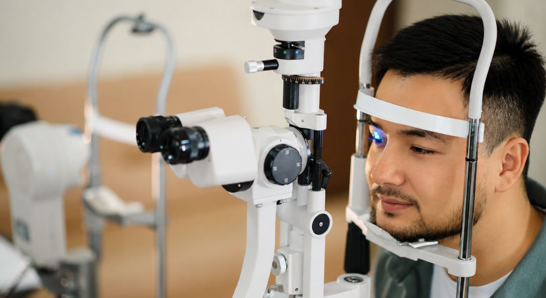
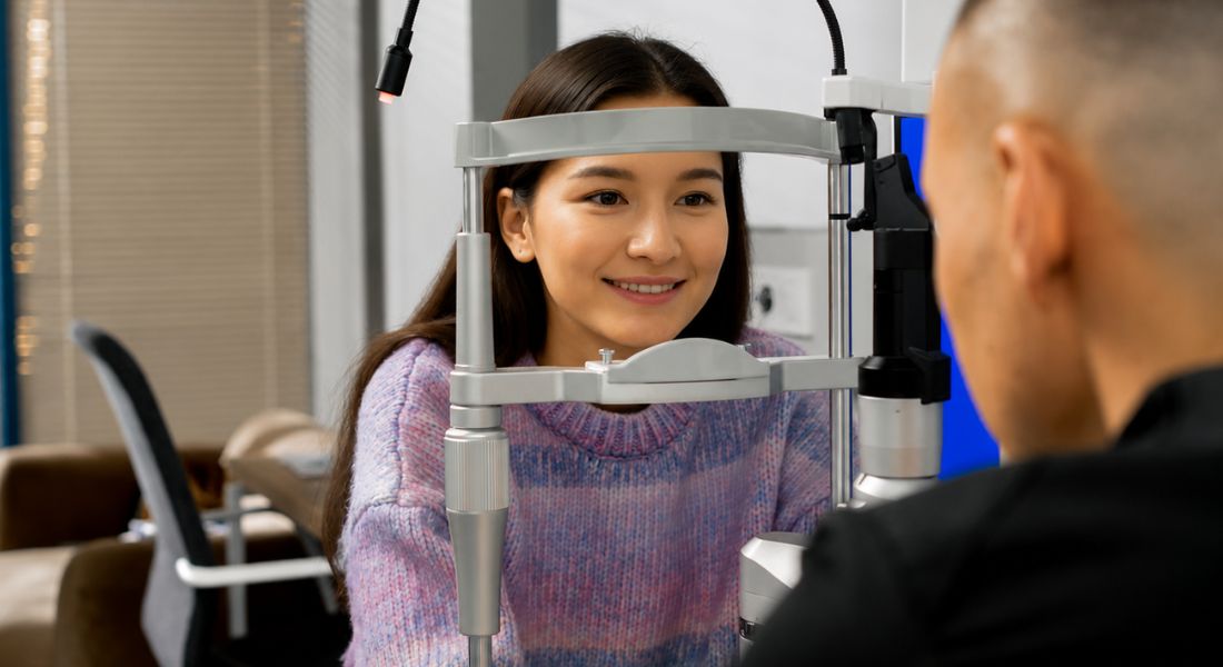
Corneal Topography and Tomography
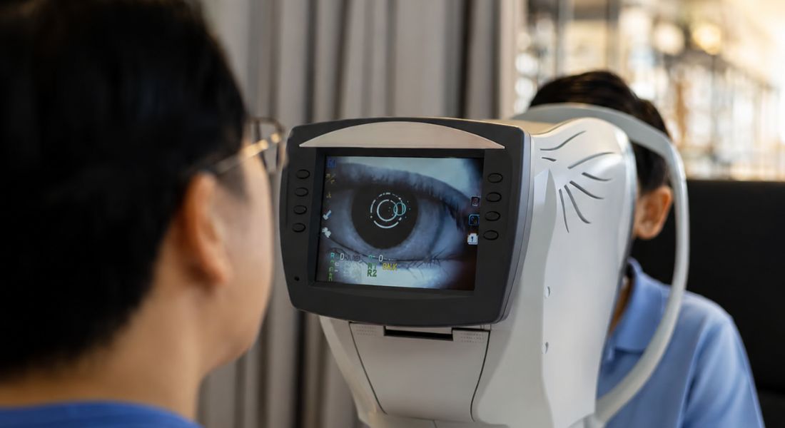
Corneal topography maps the front surface of your cornea. You may find that it helps show the curvature and any irregularities that can affect how light is focused and how clearly you see.
Corneal tomography provides a deeper three-dimensional view of the cornea, including its shape, thickness, and back surface. You might notice that this gives a more complete understanding of corneal structure than a standard sight test alone.
These scans are essential for diagnosing keratoconus and monitoring whether it is progressing over time. You may find that they can detect subtle changes that are not visible during routine eye examinations, especially in early or mild cases.
If you have risk factors or suspicious symptoms, these scans can help confirm whether keratoconus is present. You might notice that this allows for earlier diagnosis and more timely management to help protect vision.
Keratoconus Progression in Asian Patients
Keratoconus progression refers to the gradual worsening of the condition, where the cornea becomes thinner, steeper, or more irregular over time. You may find that the rate of progression can vary from person to person depending on factors such as age, eye rubbing habits, disease stage, corneal thickness, and individual biological differences.
Because some Asian patients may present at a younger age, careful monitoring can be especially important. You might notice that younger age is often associated with a longer time window for potential progression, which makes regular follow-up even more valuable in tracking changes early.
Regular corneal scans help your specialist assess whether the condition is stable or changing. You may find that these scans play a key role in deciding whether simple observation is enough or whether active treatment, such as corneal cross-linking, should be considered.
Corneal Cross-Linking
Corneal cross-linking is a treatment designed to strengthen the cornea and help slow or stop the progression of keratoconus. You may find that it is most often considered when corneal scans show clear signs that the condition is getting worse over time.
This treatment does not usually remove all existing visual distortion. Its main purpose is to stabilise the cornea and prevent further weakening, which can help reduce the risk of the condition progressing to more advanced stages.
For younger patients and those at higher risk of progression, early discussion of cross-linking can be especially important. You might notice that timely consideration of treatment can play a key role in protecting long-term vision and reducing the need for more complex interventions later.
Glasses and Contact Lens Options
In early keratoconus, glasses may still give you good functional vision, especially if the corneal changes are mild. However, as the cornea becomes more irregular, you may notice that glasses no longer provide sharp or stable vision. This is often when contact lenses become a more effective option.
- Glasses in Early Stages: In the early phase, glasses can still correct vision reasonably well, although prescriptions may change more frequently.
- Rigid Gas Permeable Lenses: These lenses help by creating a smooth optical surface over the irregular cornea, improving clarity.
- Hybrid Lenses: These combine a rigid centre with a soft outer edge to improve comfort while maintaining good vision.
- Scleral Lenses: These are larger lenses that sit over the cornea and can be particularly helpful in more irregular cases by creating a smooth, stable vision surface.
Overall, contact lenses often become more important as keratoconus progresses and glasses become less effective. The right option for you depends on how irregular your cornea is and how well you tolerate different lens types. A specialist fitting can help find the most comfortable and effective solution for your vision needs.
Advanced Keratoconus Management
If keratoconus becomes more advanced, you may find that treatment becomes more complex and may involve a combination of different specialist approaches. Some patients may need specialist contact lenses, intracorneal ring segments, laser-guided procedures in carefully selected stable cases, or in more severe situations, corneal transplant surgery.
The main aim of management is always to protect useful vision and prevent unnecessary progression. You might notice that this is why early diagnosis is so important, as it can reduce the likelihood of needing more invasive treatments later by allowing earlier monitoring and intervention.
Treatment decisions should always be based on your individual scans, symptoms, age, corneal thickness, stage of the condition, and visual needs. You may find that this personalised approach helps ensure the safest and most effective plan for preserving your vision over time.
Does Asian Background Change Treatment?
Asian background does not automatically change keratoconus treatment. You may find that treatment decisions are based primarily on your individual corneal measurements, the level of progression, your age, symptoms, and how your vision is affected.
However, research suggesting higher rates in some Asian populations may influence how carefully clinicians look for early signs during routine screening. You might notice that this can sometimes lead to earlier corneal imaging when symptoms or prescription changes appear suspicious.
The key message is that treatment is not different because of ethnicity. You may find it more accurate to think of it as a focus on earlier awareness, timely investigation, and careful monitoring based on clinical findings rather than background alone.
What This Means Before Laser Eye Surgery
Keratoconus screening is an essential part of assessment before laser eye surgery. You may find that procedures such as LASIK are not suitable if the cornea is unstable or already shows early signs of ectasia, as reshaping a weakened cornea can increase the risk of complications.
This becomes especially important if you have high astigmatism, a family history of keratoconus, or corneal scans that appear borderline. You might notice that a responsible clinic will carefully evaluate corneal shape, thickness, and overall stability before recommending any form of laser vision correction.
If keratoconus is suspected, the priority should always be corneal safety rather than proceeding with elective vision correction surgery. You may find that in such cases, further monitoring or alternative management options are considered to protect the long-term health of your eyes.
When to See a Specialist
You should consider specialist assessment if your optician mentions irregular astigmatism, corneal steepening, thinning, or a possible suspicion of keratoconus. You may find that these signs often need more detailed testing than a routine eye exam can provide, especially if your vision is not stabilising.
You should also seek advice if your vision continues to change despite updated glasses. You might notice that frequent prescription changes or ongoing blurring can be a sign that further corneal investigation is needed.
If you are looking for keratoconus treatment in London, you may want to choose a clinic that offers detailed corneal imaging, ongoing progression monitoring, advice about corneal cross-linking, and access to specialist contact lens support. This can help ensure you receive a complete and accurate assessment of your condition.
Early assessment gives you more options for management and can help protect your long-term vision. You may find that identifying changes sooner allows for closer monitoring and more timely treatment if the condition is progressing.
Future Research in Asian Patients
Future keratoconus research in Asian patients is likely to focus on several key areas, including genetics, early detection, population screening, eye rubbing habits, allergy, artificial intelligence, and predicting how quickly the condition may progress. You may find that these areas are especially important for improving earlier identification and long-term management.
More detailed studies are also needed because Asian populations are highly diverse, with differences in genetics, environment, lifestyle, and healthcare access. You might notice that research which separates different Asian subgroups could provide more accurate and meaningful insights than studies that group all Asian patients together under one category.
The overall goal of this research is earlier diagnosis, better monitoring, and more personalised treatment. You may find that improved understanding in these areas can help clinicians tailor care more effectively and reduce the risk of delayed detection or progression-related vision loss.
FAQs:
- What is keratoconus?
Keratoconus is a progressive eye condition where the cornea becomes thinner and bulges into a cone-like shape. This change affects how light enters the eye, leading to blurred vision, distortion, glare, haloes, and increasing astigmatism. It often begins in teenage years or early adulthood and can gradually worsen if not monitored. - Is keratoconus more common in Asian patients?
Some research suggests that keratoconus may be diagnosed more frequently in certain Asian populations compared with some white populations. However, “Asian” includes many different groups, and risk is not the same across all regions. The differences seen in studies may relate to genetics, environmental factors, and differences in detection rates. - Why does keratoconus vary between different Asian regions?
Prevalence varies because Asia is highly diverse, including South Asian, East Asian, Southeast Asian, West Asian, and Central Asian populations. Studies show that rates differ significantly between these regions. Differences in diagnostic methods, healthcare access, and environmental factors also influence reported prevalence. - What are the early signs of keratoconus?
Early signs include frequent changes in glasses prescription, increasing astigmatism, blurred or distorted vision, ghosting around letters, glare, and difficulty seeing at night. These symptoms may develop gradually, so they are often missed without detailed corneal imaging. - How is keratoconus diagnosed?
Keratoconus is diagnosed using corneal topography and tomography, which map the shape and thickness of the cornea in detail. These scans can detect early or subtle changes before vision becomes significantly affected, making them essential for early diagnosis and monitoring. - Does eye rubbing increase the risk?
Yes. Eye rubbing is one of the strongest modifiable risk factors linked with keratoconus. Repeated rubbing places mechanical stress on the cornea and may contribute to progression. Eye rubbing is often linked with allergies, dryness, or irritation, so treating the underlying cause is important. - Do allergies play a role in keratoconus?
Yes, allergies are commonly associated with keratoconus because they often lead to itchy eyes and rubbing. Conditions such as hay fever, eczema, and allergic conjunctivitis are frequently reported in patients. While allergies do not directly cause keratoconus, they may contribute indirectly through chronic eye rubbing. - How is keratoconus treated or managed?
Treatment depends on severity. In early stages, glasses may still be effective. As it progresses, specialist contact lenses such as rigid, hybrid, or scleral lenses may be needed. If the condition is progressing, corneal cross-linking may be used to strengthen the cornea and help stop further deterioration. - Can keratoconus be prevented?
There is no guaranteed way to prevent keratoconus, especially when genetic factors are involved. However, reducing eye rubbing, managing allergies, and having regular eye examinations can help with early detection and may reduce the risk of progression. - When should Asian patients see a specialist?
You should seek specialist assessment if you experience frequent prescription changes, increasing astigmatism, blurred or distorted vision, or if glasses no longer fully correct your sight. A family history of keratoconus or suspicion of corneal irregularity are also important warning signs that require corneal imaging and proper evaluation.
Final Thoughts: Understanding Keratoconus Awareness and Early Detection in Asian Patients
Keratoconus research suggests that some Asian populations may show higher reported prevalence and earlier presentation compared with other groups, but the most important message is not about ethnicity alone. It is about recognising symptoms early, identifying risk factors such as eye rubbing and family history, and ensuring timely access to detailed corneal imaging. When keratoconus is detected early, outcomes are generally much better because progression can often be monitored or stabilised before significant vision loss occurs.
The key focus in modern eye care is individual assessment rather than assumptions based on background. Regular eye checks, awareness of subtle vision changes, and early referral for corneal topography or tomography can make a significant difference. With appropriate monitoring and treatments such as corneal cross-linking and specialist contact lenses, most patients can maintain good functional vision. If you are exploring whether keratoconus treatment in London may be suitable for your needs, get in touch with us at Eye Clinic London to schedule your consultation.
References:
- Georgiou, T., Funnell, C.L., Cassels-Brown, A. and O’Conor, R. (2004) Influence of ethnic origin on the incidence of keratoconus and associated atopic disease in Asian and white patients, 18, pp. 379–383. Available at: https://pubmed.ncbi.nlm.nih.gov/15069434/
- Gomes, J.A.P., Rodrigues, P.F. and Lamazales, L.L. (2022) Keratoconus epidemiology: a review, Saudi Journal of Ophthalmology, 36(1), pp. 3–6. Available at: https://www.mdpi.com/2079-4983/5/3/111
- Grunauer-Kloevekorn, C. and Duncker, G.I. (2006) Keratoconus: epidemiology, risk factors and diagnosis, Available at: https://pmc.ncbi.nlm.nih.gov/articles/PMC3775068
- Khor, W.B., Wei, R.H., Lim, L., Chan, C.M. and Tan, D.T. (2011) Keratoconus in Asians: demographics, clinical characteristics and visual function in a hospital-based population, Clinical & Experimental Ophthalmology, 39(4), pp. 299-307. Available at: https://pubmed.ncbi.nlm.nih.gov/21070542/
- Hassan, Z., et al. (2020) The enigma of environmental factors in keratoconus, 9(6), pp. 549–556. Available at: https://www.sciencedirect.com/science/article/pii/S2162098923001627