

Keratoconus in Black Patients: What Does the Research Show?
Keratoconus is a condition where the cornea becomes thinner, weaker, and gradually takes on a more cone-like shape over time. You may find that because the cornea is the clear front part of the eye, these structural changes can significantly affect how light is focused, leading to blurred vision, distortion, glare, haloes, and increasing astigmatism.
Research has identified differences in keratoconus prevalence, diagnosis, severity, and access to care between different populations. You might notice that some recent US studies suggest keratoconus may be more common in Black patients compared with certain other groups, and that diagnosis may sometimes be delayed in these patients due to a range of healthcare, access, and referral-related factors.
You may find it important to understand that these findings do not mean race alone causes keratoconus. Instead, they reflect a combination of biological variation, environmental influences, healthcare access, and differences in how early the condition is detected and managed across populations.
This does not mean every Black patient is at high risk. You may find it more accurate to view it as a reminder that awareness is important, especially if you notice symptoms such as frequent prescription changes, strong astigmatism, distorted vision, eye rubbing, allergies, or a family history of keratoconus, all of which should be properly assessed rather than ignored.
What Is Keratoconus?
Keratoconus affects the cornea, which is the clear front surface of your eye and normally has a smooth, rounded shape. You may find that in keratoconus, the cornea gradually becomes thinner and begins to bulge forward into a cone-like shape.
This change in shape affects how light enters and is focused inside the eye. You might notice that vision becomes blurred, doubled, ghosted, or distorted, and it may become increasingly difficult to correct fully with standard glasses as the condition progresses.
The condition often begins during the teenage years or early adulthood. You may find that this is why early eye checks and regular monitoring are important, as they can help detect changes sooner and support better long-term vision care.
Why Black Patients Are Discussed in Keratoconus Research
Black patients are often discussed in keratoconus research because recent studies have reported differences in both prevalence and clinical presentation across racial and ethnic groups. You may find that a 2024 US-based study reported higher keratoconus prevalence in Black and Hispanic populations compared with White and Asian populations.
Another large US study also found that keratoconus prevalence was higher in the Black population, particularly among female individuals, and that diagnosis was often delayed in these patients. You might notice that this highlights not only differences in how common the condition appears, but also how and when it is identified in clinical practice.
These findings do not mean that race alone causes keratoconus. You may find it more accurate to understand them as showing that multiple factors biological, environmental, social, diagnostic, and healthcare access-related can all influence risk and detection patterns.
What the Research Says About Prevalence
Keratoconus prevalence can vary widely depending on the country, age group, diagnostic method, and study design. You may find that this is because older studies often relied on clinical diagnosis alone, while newer research may use more advanced tools such as imaging, insurance databases, or electronic health records to identify cases more accurately.
A 2024 article on socioeconomic and demographic disparities notes that keratoconus is most common in people aged 18 to 39 years and has been reported with higher prevalence in Black and Hispanic populations in the United States. You might notice that these findings highlight both age-related risk patterns and differences observed across population groups.
For patients, the practical message is simple. If symptoms are present, such as changing vision, increasing astigmatism, or distortion, the cornea should be properly examined with appropriate testing rather than assuming it is only a glasses prescription issue.
Why Prevalence Numbers Can Differ
Prevalence numbers for keratoconus can vary significantly between studies because different research groups use different tools, definitions, and diagnostic thresholds. You may find that mild or early keratoconus can be missed if advanced imaging like corneal topography or tomography is not used.
With modern imaging techniques, clinicians are able to detect early or very subtle disease that may not be obvious during a standard eye examination. You might notice that this can make newer studies appear to report higher prevalence rates compared with older research that relied more on symptoms or basic clinical findings.
A worldwide epidemiology review explains that keratoconus prevalence differs across regions and populations, partly due to variations in diagnostic criteria and screening methods. You may find that this makes direct comparisons between studies more complex, as results depend heavily on how keratoconus is defined and detected.
Delayed Diagnosis in Black Patients
Delayed diagnosis is an important concern in keratoconus. You may find that if the condition is identified late, the cornea can already be more irregular or more advanced, which can make management more challenging.
A US prevalence and burden study reported that keratoconus diagnosis is often delayed in Black patients. You might notice that this does not point to a single cause, but rather reflects a combination of factors such as access to care, frequency of eye examinations, referral timing, and availability of specialist imaging.
This matters because earlier diagnosis can allow earlier monitoring and treatment. You may find that identifying keratoconus sooner gives more opportunity to intervene, including considering corneal cross-linking if there are signs that the condition is progressing.
Severity at Presentation
Some studies suggest that Black patients may present with more advanced keratoconus compared with non-Black patients. You may find that this is not only related to biological factors, but also influenced by delayed diagnosis, reduced access to specialist corneal imaging, differences in referral pathways, and wider healthcare inequalities.
An ARVO abstract on racial and ethnic disparities reported that Black patients often presented with more advanced keratoconus than non-Black patients in the authors’ clinical setting. You might notice that such findings highlight the importance of improving early access to eye care and ensuring timely use of diagnostic scans when symptoms first appear.
This is why symptoms such as worsening vision, frequent prescription changes, or increasing astigmatism should be investigated early. You may find that early assessment can help detect keratoconus sooner, allowing for closer monitoring and treatment before the condition progresses to a more advanced stage.
Visual Impairment and Race
Research has also examined whether keratoconus is associated with different patterns of visual impairment across racial groups. You may find that a 2023 study on racial variation in keratoconus patients discussed differences in presentation and highlighted the importance of considering both race and sex when analysing keratoconus data.
This type of research is important because keratoconus does not affect every person in the same way. You might notice that some patients are diagnosed early and remain stable for many years, while others experience faster progression or present later in the course of the disease.
You may find that these differences are influenced by a combination of biological, environmental, and healthcare-related factors rather than a single cause. This is why researchers continue to study large and diverse populations to better understand patterns of disease.
Better data can help clinicians identify which patients may need closer monitoring. You might notice that this ultimately supports earlier detection, more tailored follow-up, and improved long-term visual outcomes for people at higher risk.
The Role of Genetics
Genetics can play a role in keratoconus, although it is usually a complex condition rather than being caused by a single gene. Family history is an important risk factor, and it can increase the likelihood of developing the condition. However, not everyone with a family history will go on to develop keratoconus.
- Family History Matters: If a parent, sibling, or close relative has keratoconus, your own risk may be higher.
- Complex Genetic Influence: Keratoconus is not linked to one specific gene in most cases, but rather a combination of genetic and environmental factors.
- Risk Across All Ethnic Groups: Increased risk due to family history applies regardless of ethnicity.
- Importance of Sharing History: Mentioning family history during an eye test can help your clinician decide whether closer monitoring or corneal imaging is needed.
Overall, genetics is one important factor in keratoconus risk, but it is not the only one. Early awareness of family history can support earlier detection and better monitoring, especially in younger patients with changing vision.
Corneal Thickness and Ancestry
Research has explored possible links between ancestry and corneal thickness, and you may find that this is one reason keratoconus risk is studied across different populations. A Kaiser Permanente genetics report described findings suggesting that people with greater African ancestry were more likely, on average, to have thinner corneas, which may be relevant when considering susceptibility to certain corneal conditions.
However, this does not mean that all people with African ancestry have thin corneas or will develop keratoconus. You might notice that this type of research is focused on population-level patterns rather than individual outcomes, and there is always significant variation from person to person.
You may find it important to understand that individual corneal scans are far more reliable than assumptions based on background or ancestry. These scans give a precise measurement of your own corneal structure and are what clinicians use to assess real risk.
Overall, this research helps explain why scientists are interested in how corneal characteristics may differ between populations, but it does not replace personalised clinical assessment in diagnosing or managing keratoconus.
Eye Rubbing and Allergy

Eye rubbing is one of the most important modifiable risk factors linked with keratoconus. You may find that repeated rubbing places mechanical stress on the cornea, and over time this can contribute to weakening or changes in its shape, especially in people who are already more susceptible.
Many people rub their eyes because of conditions like allergy, eczema, asthma, hay fever, dry eye, or general irritation. You might notice that this becomes a habit, especially when symptoms flare up, but treating the underlying itch or discomfort is usually far more helpful than continuing to rub the eyes.
If you experience itchy eyes, redness, watering, or seasonal allergies, it is worth discussing treatment with an eye specialist or optometrist. You may find that managing these symptoms properly can reduce the urge to rub your eyes and help protect your long-term corneal health.
Symptoms Black Patients Should Not Ignore
You should not ignore frequent changes in your glasses prescription, especially if you notice your astigmatism is increasing. You may find that this can be one of the early signs that your cornea is changing shape and needs further checking.
You should also pay attention if your vision remains blurred even after getting new glasses. Other symptoms you might notice include ghosting around letters, glare, haloes around lights, light sensitivity, eye strain, or difficulty driving at night. In some cases, one eye may also seem noticeably worse than the other.
These symptoms do not always mean you have keratoconus, but you may find that they are important enough to warrant a proper corneal assessment. Early scanning can help confirm what is happening and guide the right next steps for your eye health.
Why Teenagers and Young Adults Need Attention
Keratoconus often begins in adolescence or early adulthood, which is an important time because the condition may have many years to progress. In younger patients, changes in the cornea can sometimes happen more quickly, so early recognition is key. This is why symptoms in teenagers and young adults should always be taken seriously and assessed properly.
- Early Onset Is Common: Keratoconus frequently starts during the teenage years or early adulthood when the cornea is still changing.
- Faster Progression Risk: Younger patients may experience faster progression, making early monitoring especially important.
- Warning Signs Matter: Rapidly changing glasses, increasing astigmatism, eye rubbing, or a family history should raise suspicion.
- Routine Tests May Not Be Enough: Standard sight tests may miss early changes, so corneal imaging may be needed for accurate diagnosis.
Overall, early attention in teenagers and young adults can make a significant difference in protecting long-term vision. When keratoconus is identified early, there is a better chance of monitoring progression and considering treatments before the condition becomes more advanced. Timely referral to a specialist helps ensure the most appropriate care at the right stage.
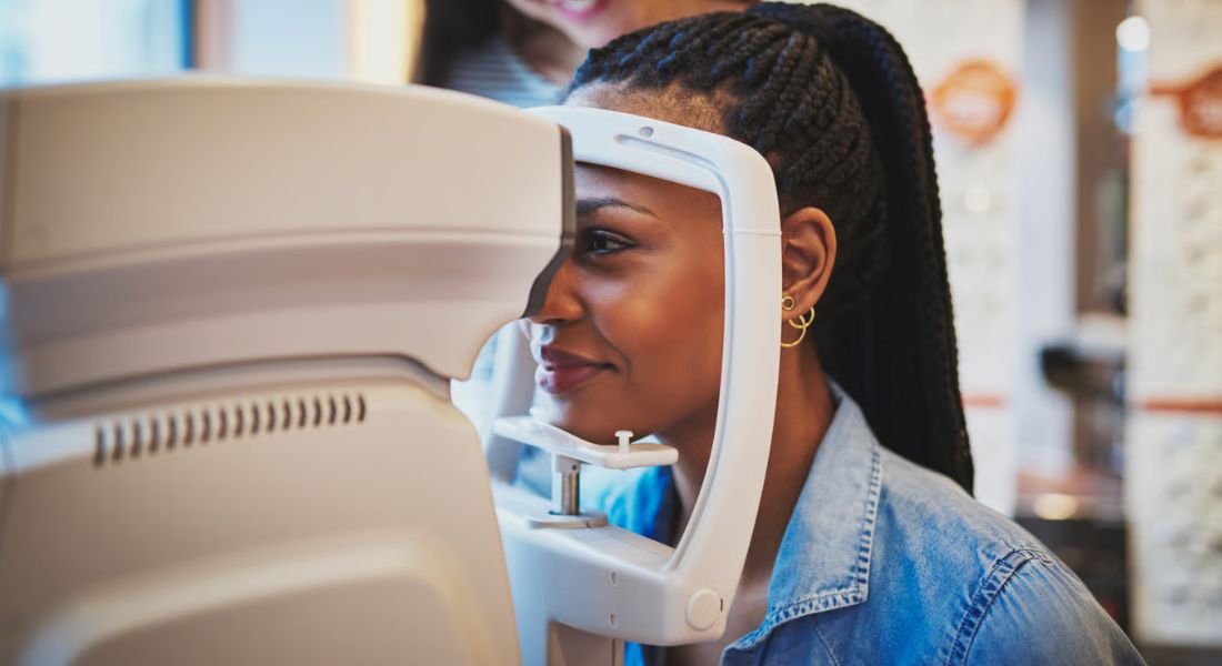
Corneal Topography and Tomography
Corneal topography maps the front surface of your cornea. You may find that it helps identify subtle irregularities in corneal shape that can affect how light is focused and how clearly you see.
Corneal tomography provides a more detailed three-dimensional view of the cornea. It can show changes in thickness and the back surface shape, giving a more complete understanding of corneal structure than a standard eye test alone.
These scans are essential for detecting early keratoconus and monitoring whether it is progressing over time. You might notice that they can pick up changes even before symptoms become obvious during a routine eye examination.
For patients with symptoms or known risk factors, these scans can help confirm whether keratoconus is present. You may find that this allows for earlier diagnosis and more timely management to help protect vision.
Corneal Cross-Linking
Corneal cross-linking is a treatment used to strengthen the cornea and help slow or stop the progression of keratoconus. You may find that it does not usually remove all existing visual distortion, but it can be very effective in preventing the condition from getting worse over time.
This treatment is especially important when keratoconus is diagnosed early. If progression is confirmed on scans, you might be advised that cross-linking can reduce the risk of more advanced changes developing later, which may help avoid more complex treatment in the future.
The decision to proceed with cross-linking depends on several factors, including your age, changes seen on corneal scans, corneal thickness, stage of the disease, and overall eye health. You may find that this careful assessment helps ensure the treatment is only recommended when it is truly beneficial for your situation.
Glasses and Contact Lens Options

In early keratoconus, you may find that glasses can still provide useful vision and help you manage day-to-day activities. However, as the cornea becomes more irregular over time, glasses may start to become less effective in giving you clear and stable vision.
At this stage, specialist contact lenses are often recommended. You might be offered options such as rigid gas permeable lenses, hybrid lenses, or scleral lenses, depending on your corneal shape and level of visual distortion.
Scleral lenses can be especially helpful for irregular corneas because they create a smooth optical surface over the eye. You may find that this helps improve comfort and clarity by reducing the visual distortion caused by the uneven corneal shape.
Advanced Keratoconus Treatment
If keratoconus becomes advanced, you may find that treatment becomes more complex and may involve a combination of different approaches depending on how the cornea has changed. Some patients may need specialist contact lenses, intracorneal ring segments, laser-guided procedures in selected stable cases, or in more severe situations, corneal transplant surgery.
The main aim of treatment is always to protect your vision and prevent further unnecessary progression. You might notice that this is why earlier diagnosis is so important, as it often allows for more treatment options and better long-term control of the condition.
Advanced treatment decisions should always be guided by detailed corneal scans and specialist assessment. You may find that careful evaluation helps ensure the treatment plan is tailored to your specific corneal shape, level of progression, and visual needs.
Does Black Background Change Treatment?
A Black background does not automatically change how keratoconus is treated. Treatment decisions are based on your individual corneal shape, thickness, rate of progression, age, symptoms, vision, and overall eye health. The same clinical principles apply regardless of ethnicity, and the focus remains on protecting vision and slowing progression.
- Treatment Is Individualised: Your management plan depends on your eye measurements and disease behaviour, not your racial background.
- Earlier Detection May Be Encouraged: If research suggests higher risk or later diagnosis in some groups, clinicians may be more alert to early signs and consider earlier corneal scans.
- Symptoms Still Guide Referral: Changes in vision, increasing astigmatism, or poor visual quality remain key reasons for further investigation.
- Access and Awareness Matter: Ensuring timely referral and specialist assessment is important for all patients, especially where delayed diagnosis is more likely.
Overall, Black background does not change the treatment itself, but it may influence awareness and vigilance around diagnosis. The goal is earlier identification, fair access to care, and timely treatment when needed. This helps ensure that outcomes are based on disease severity rather than timing of diagnosis.
Healthcare Access and Diagnosis
Healthcare access can play an important role in when keratoconus is diagnosed. You may find that if someone has fewer routine eye examinations, limited access to corneal imaging, or delayed referral to a specialist, the condition may only be identified at a later stage.
A 2024 study examining socioeconomic and demographic disparities in keratoconus highlighted the importance of recognising that diagnosis and management are not always equal across different groups. You might notice that this means factors such as access to care and healthcare awareness can influence when the condition is picked up.
This is important because early treatment can make a significant difference to long-term outcomes for many patients. You may find that timely diagnosis allows for closer monitoring, earlier intervention, and better protection of vision over time.
What This Means for Families
If keratoconus runs in your family, you may find it helpful for close relatives to have regular eye checks, especially teenagers and young adults. You might also notice that mentioning a family history of keratoconus during routine optician appointments can help guide more careful examination and earlier screening.
Parents should pay attention if a child frequently rubs their eyes, struggles to see clearly with glasses, complains of ghosting, or needs repeated changes in prescription. You may find that these signs do not confirm keratoconus, but they are important enough to justify a proper eye assessment.
Family awareness can play a key role in earlier detection of keratoconus. You might notice that when relatives are informed and observant, the condition can be identified sooner, which helps with timely monitoring and treatment if needed.
When to See a Specialist

You should consider specialist assessment if your optician mentions irregular astigmatism, corneal steepening, thinning, or a possible suspicion of keratoconus. You may also find it important to seek advice if your vision keeps changing even after you have updated glasses, as this can be a sign that something more detailed needs to be checked.
If you are looking for keratoconus treatment in London, you may want to choose a clinic that can offer detailed corneal imaging, ongoing monitoring, clear advice about cross-linking, and access to specialist contact lens support. This can help ensure you get a complete understanding of your eye health rather than just a basic vision check.
The earlier you understand what is happening, the easier it is to protect your vision in the long term. You may find that early assessment allows for more options, better planning, and closer monitoring to help slow progression if needed.
Future Research in Black Patients
Future research in Black patients with keratoconus is likely to focus on several important areas, including genetics, corneal structure, early diagnosis, treatment access, disease progression, and the impact of healthcare disparities. You may find that a key reason for this focus is that older keratoconus research has not always included all populations equally, so clearer and more inclusive data is still needed.
Researchers are also expected to study how modern tools such as advanced imaging, artificial intelligence, and population-based screening can improve earlier detection. You might notice that this is particularly important in settings where keratoconus is currently diagnosed later than ideal, which can affect treatment outcomes.
These developments are aimed at improving fairness and accuracy in eye care. You may find that the long-term goal is to ensure earlier diagnosis and more effective management of keratoconus for every patient, regardless of background, so that vision can be protected as much as possible.
FAQs:
- What is keratoconus?
Keratoconus is a progressive eye condition where the cornea becomes thinner and gradually bulges into a cone-like shape. This changes how light enters the eye, leading to blurred vision, ghosting, glare, and increasing astigmatism. In many cases, glasses help only in the early stages, and more specialised treatment may be needed as the condition progresses. - Is keratoconus more common in Black patients?
Some research, particularly from the United States, suggests that keratoconus may be diagnosed more frequently in Black and Hispanic populations compared with some other groups. However, this does not mean race alone causes the condition. The differences are likely influenced by a combination of genetics, environmental factors, healthcare access, and how early the disease is detected. - Why might keratoconus be diagnosed later in Black patients?
Studies suggest that keratoconus may sometimes be diagnosed later in Black patients due to a combination of factors, including reduced access to specialist eye imaging, delayed referrals, and differences in routine eye care uptake. As a result, some patients may already have more advanced disease by the time they are diagnosed, which can affect treatment outcomes. - What are the early warning signs of keratoconus?
Early signs include frequent changes in glasses prescription, increasing astigmatism, blurred or distorted vision, ghosting around letters, and difficulty seeing clearly at night. Some patients also notice eye strain or sensitivity to light. These symptoms can develop gradually, which is why early corneal scans are important. - How is keratoconus diagnosed?
Keratoconus is diagnosed using advanced corneal imaging such as topography and tomography. These scans measure the shape, curvature, and thickness of the cornea in detail. This allows specialists to detect early or subtle changes that may not be visible during a standard eye examination. - Does eye rubbing increase the risk of keratoconus?
Yes. Eye rubbing is one of the strongest modifiable risk factors linked with keratoconus progression. Repeated rubbing can weaken the cornea over time, especially in individuals who already have genetic susceptibility. Many people rub their eyes due to allergies or irritation, so treating the underlying cause is important. - Does Black ethnicity change the symptoms of keratoconus?
No, the symptoms of keratoconus are generally the same across all ethnic groups. Patients typically experience blurred vision, distortion, glare, haloes, and difficulty with night vision. The main differences reported in research relate more to timing of diagnosis and severity at presentation rather than symptom type. - Can keratoconus be treated or slowed down?
Yes. While keratoconus cannot usually be reversed, its progression can often be slowed or stabilised. Corneal cross-linking is a key treatment that strengthens the cornea and helps stop further deterioration. Glasses and specialist contact lenses may also improve vision depending on the stage of the condition. - What role do genetics and family history play?
Family history is an important risk factor. If a close relative has keratoconus, your risk may be higher, regardless of ethnicity. Genetic factors may influence corneal strength, but keratoconus is usually caused by a combination of inherited and environmental influences rather than a single gene. - When should someone seek specialist eye care?
You should seek specialist assessment if your vision keeps changing, astigmatism is increasing, or glasses no longer provide clear vision. A family history of keratoconus, frequent eye rubbing, or suspected corneal irregularity are also important warning signs. Early referral allows for timely scanning and better long-term protection of vision.
Final Thoughts: Improving Awareness and Early Diagnosis of Keratoconus in Black Patients
Keratoconus research highlights that differences in prevalence, severity, and timing of diagnosis may be seen across populations, including Black patients. However, the most important message is not about race itself, but about ensuring early recognition of symptoms and timely access to proper corneal assessment. When keratoconus is detected early, options such as monitoring and corneal cross-linking can significantly reduce the risk of long-term vision loss.
In everyday clinical practice, what truly matters is how quickly changes in vision are investigated rather than background alone. Symptoms such as frequent prescription changes, increasing astigmatism, blurred or distorted vision, and difficulty with night vision should always be checked with detailed corneal imaging. With modern diagnostic techniques and improved awareness, keratoconus can often be identified earlier and managed more effectively than in the past. If you are considering keratoconus treatment in London and want to know if it’s the right option, you’re welcome to reach out to us at Eye Clinic London to book a consultation.
References:
- Singh, R.B. et al. (2024) Prevalence and economic burden of keratoconus in the United States, American Journal of Ophthalmology. Available at: https://pubmed.ncbi.nlm.nih.gov/37951332/
- Dreyer, E.B. et al. (2023) Racial variation in visual impairment of keratoconus patients at presentation. Available at: https://pmc.ncbi.nlm.nih.gov/articles/PMC10709529/
- Holland, E.J. et al. (2015) Association between sociodemographic factors and keratoconus. Available at: https://pubmed.ncbi.nlm.nih.gov/26707415/
- Lucas, S.E. and Burdon, K.P. (2013) Epidemiology of keratoconus, Clinical and Experimental Ophthalmology. Available at: https://pmc.ncbi.nlm.nih.gov/articles/PMC3775068/
- Pederzolli, M., Procopio, F., Tombolini, B., Marra, S., De Micheli, M., Bandello, F. and Ferrari, G. (2025) Keratoconus: the local manifestation of a systemic disease? Journal of Clinical Medicine, 14(13), 4587. Available at: https://www.mdpi.com/2077-0383/14/13/4587