

How Is Your Cornea Mapped Before Laser Eye Surgery? (Topography Explained)
When you first start exploring laser eye surgery, it is easy to assume the process begins and ends with the laser itself. In reality, a significant amount of preparation takes place long before any treatment is carried out. This preparation is essential for ensuring that your eyes are properly understood and that the procedure is both safe and effective. It is this groundwork that guides every decision your surgeon makes.
One of the most important parts of this preparation is corneal mapping, also known as corneal topography. This involves scanning your eye in great detail to create a precise map of its surface and structure. The map shows the shape, curvature, and subtle variations of your cornea. It provides information that cannot be gathered through a standard eye examination alone.
Corneal topography allows your surgeon to see your eye in a much more detailed and accurate way. It helps identify how light is currently entering your eye and where any irregularities may be affecting your vision. This level of detail is crucial for planning how the laser will reshape your cornea. It ensures that treatment is tailored specifically to your individual eye structure.
If you have ever wondered how surgeons decide exactly how to treat your vision, this is where the process begins. The information gathered from corneal mapping is used alongside other measurements to build a complete picture of your eye health. This includes factors like corneal thickness and residual stromal bed calculations. Together, these details allow your surgeon to design a treatment plan that is precise, safe, and fully personalised.
Why Corneal Mapping Is So Important
Your cornea is not perfectly uniform, even though it may look smooth and clear to the naked eye. In reality, its surface contains tiny variations in shape, thickness, and curvature. These differences are completely normal, but they still play an important role in how your vision is formed. Even small irregularities can influence visual quality.
These variations matter because they affect how light enters your eye and focuses on the retina. Your vision depends on light being accurately directed to create a clear image. If there are subtle imperfections in the cornea’s shape, this can lead to blurred or distorted vision. That is why these small details are taken very seriously during assessment.
When planning laser eye surgery, your surgeon needs a complete and precise understanding of your cornea. Corneal mapping provides this by creating a detailed image of its exact structure and curvature. This allows treatment to be fully tailored to your eye rather than using a general approach. Without this level of detail, achieving safe and predictable results would not be possible.
What Is Corneal Topography?
Corneal topography is one of the most important diagnostic tools used during a laser eye surgery assessment. It provides a highly detailed map of the surface of your cornea, helping surgeons understand its shape and structure. This information is essential for planning safe and accurate treatment. By analysing the cornea in detail, surgeons can detect irregularities that may not be visible through standard eye tests.
- A Detailed 3D Map of the Cornea: Corneal topography works by creating a three-dimensional map of the corneal surface. It functions much like a 3D scanner, capturing thousands of data points across the eye. This allows surgeons to see the exact shape and contour of the cornea in great detail.
- Light Projection and Reflection Analysis: The device projects a pattern of light onto the surface of the eye and measures how it reflects back. These reflections are analysed by specialised computer software to build an accurate representation of the cornea. This process is quick, precise, and completely non-invasive.
- Colour-Coded Mapping System: The results are displayed as a colour-coded map that represents different curvature levels. Steeper areas are shown in warmer colours, flatter areas in cooler tones, and irregular regions in contrasting shades. This visual system makes it easier for surgeons to quickly interpret corneal shape and identify potential concerns.
- Detecting Irregularities and Planning Surgery: Corneal topography helps identify subtle irregularities that could affect surgical outcomes. These may include uneven surfaces or early signs of corneal conditions. By understanding these details, surgeons can choose the safest and most suitable procedure for each patient.
In conclusion, corneal topography plays a vital role in ensuring laser eye surgery is both safe and effective. It provides a clear, detailed view of the cornea’s shape that cannot be achieved through basic examination alone. The test is quick, painless, and highly informative for surgical planning. Ultimately, it helps guide surgeons in delivering personalised and precise treatment outcomes.
What Is Corneal Tomography?
While corneal topography focuses mainly on the surface of your cornea, tomography goes one step further by looking deeper into its overall structure. Instead of only showing the outer shape, it builds a complete view of the cornea from front to back. This gives your surgeon a much more detailed understanding of how your eye is formed. It is an important part of modern laser eye surgery assessment.
Corneal tomography creates a 3D image of your entire cornea, including both the surface and internal layers. This allows specialists to see how thick your cornea is in different areas and how its internal shape is structured. Because it examines deeper layers, it can detect issues that may not be visible on surface scans alone. This makes it especially useful for identifying hidden irregularities early.
This advanced imaging also measures important factors such as corneal thickness, internal curvature, structural integrity, and subtle abnormalities. These details help your surgeon understand how strong and stable your cornea is before any treatment is planned. When combined with corneal topography, it provides a full and accurate picture of your eye. Together, these tools ensure your treatment is both safe and precisely tailored to you.
The Difference Between Topography and Tomography
It is easy to confuse corneal topography and tomography, but they actually serve different purposes in your eye assessment. Both are important for laser eye surgery, yet they focus on different aspects of your cornea. Understanding the difference helps you see how detailed your pre-surgery evaluation really is. Each test contributes a unique layer of information.
Corneal topography is like looking at the surface map of a landscape. It shows the outer shape of your cornea, including its curves, elevations, and depressions. This helps your surgeon understand how the surface of your eye affects your vision. It focuses entirely on the external structure.
Corneal tomography, on the other hand, goes deeper by showing what lies beneath the surface as well. It creates a 3D image of your cornea, including internal layers and thickness distribution. This is similar to looking inside the same landscape rather than just at its surface. It helps detect hidden issues that topography alone may miss.
How the Mapping Process Works Step by Step
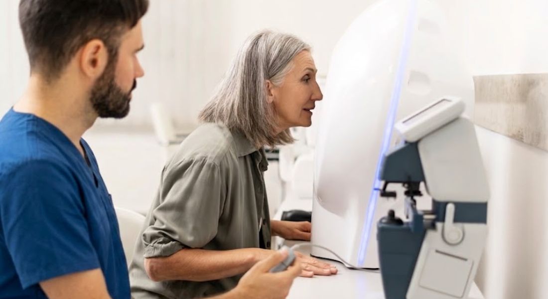
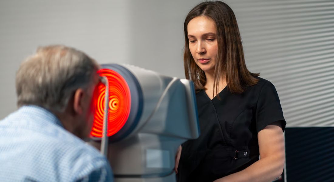
When you attend your pre-surgery consultation, corneal mapping is usually one of the first diagnostic steps. It helps your surgeon gather detailed information about your eye before any treatment is planned. The process is quick, painless, and completely non-invasive. It is designed to give a highly accurate picture of your cornea.
First, you will be seated in front of a scanning machine and asked to focus on a small light target. This helps keep your eye steady while the images are taken. The machine then captures a rapid series of detailed images of your eye within seconds. Even if you blink or move slightly, the process is still safe and completed very quickly.
Once the scan is finished, the information is processed by specialised software. This creates detailed colour maps and 3D models of your cornea, showing its shape and structure. Your surgeon then reviews these results carefully to decide the most suitable treatment plan for you.
What Surgeons Look for in Corneal Maps
Corneal mapping is far more than just producing detailed colour images of the eye. It is a vital diagnostic tool that gives surgeons a deeper understanding of corneal health and structure. These maps help identify subtle patterns that cannot always be detected through standard eye examinations. By carefully analysing this information, surgeons can make safer and more accurate decisions about surgery suitability.
- Corneal Symmetry: One of the main things surgeons assess is how symmetrical the cornea is. A healthy cornea is generally smooth and evenly shaped, while asymmetry may suggest underlying issues. Even minor imbalances can influence how light is focused and how well surgical correction will work.
- Irregular Curvature Patterns: Surgeons closely examine the curvature of the cornea to detect any irregularities. Uneven or distorted areas may indicate conditions that could affect surgical outcomes. Identifying these patterns early helps prevent complications and ensures the most appropriate procedure is chosen.
- Thickness Distribution: Corneal maps also show how thickness varies across different areas of the cornea. This is important because not all parts of the cornea are equally strong or suitable for tissue removal. Uneven thickness distribution may limit certain surgical options or require alternative techniques.
- Early Signs of Corneal Conditions: Detailed mapping can reveal early indicators of conditions such as keratoconus or other structural abnormalities. These issues may not yet be noticeable to the patient but can significantly impact surgical safety. Early detection allows surgeons to avoid unsuitable procedures and recommend safer alternatives.
- Overall Shape Stability: Surgeons also assess whether the cornea maintains a stable and consistent shape. Changes or instability over time can affect long-term surgical outcomes. A stable corneal structure is generally a key requirement for proceeding with laser eye surgery.
In conclusion, corneal mapping provides surgeons with critical insights that go far beyond surface-level examination. It helps them evaluate symmetry, structure, thickness, and overall corneal health in great detail. Even small irregularities can influence the final decision about surgery eligibility. Ultimately, this step is essential for ensuring that any treatment plan is both safe and precisely tailored to the individual’s eyes.
Why Corneal Mapping Helps Ensure Safety
Safety is the number one priority in laser eye surgery, and corneal mapping plays a direct role in making sure this is achieved. It provides your surgeon with a detailed understanding of your cornea before any treatment begins. This helps reduce uncertainty and ensures every decision is based on accurate information. It is a key step in safe surgical planning.
By identifying irregularities or weaknesses in your cornea, mapping allows your surgeon to determine whether a procedure is suitable for you. It can highlight subtle issues that may not be visible during a basic eye examination. This means potential risks can be spotted early and taken into account. As a result, your treatment plan becomes much more precise and personalised.
If tomography or topography shows signs of corneal instability, certain laser procedures may be avoided. In such cases, safer alternatives can be recommended to protect your eye health. This careful approach helps prevent complications and supports long-term stability. Ultimately, corneal mapping ensures that your treatment is both safe and well-suited to your eyes.
How Mapping Affects Your Treatment Plan

The results of your corneal scans are a key part of creating your personalised treatment plan. Once the mapping is complete, your surgeon uses this information to understand your eye in detail before making any decisions. This ensures that every recommendation is based on precise measurements rather than estimates. It is an essential step in safe and effective planning.
No two eyes are the same, which means no two laser treatments should be identical. Your cornea has its own unique shape, thickness, and structure that must all be considered carefully. Corneal mapping highlights these differences clearly so your surgeon can tailor the treatment specifically to you. This helps ensure that your procedure matches your individual eye profile.
The mapping data helps guide several important decisions, including which procedure is most suitable, how much tissue needs to be removed, and what laser settings should be used. In some cases, it may even show that surgery is not the safest option for you. This level of precision ensures that your treatment is both safe and fully personalised to your eyes.
Detecting Conditions That You Cannot Feel or See
One of the most important benefits of corneal mapping is its ability to detect hidden conditions in your eye. Many corneal issues do not cause obvious symptoms in the early stages, which means you might not notice anything unusual. Your vision can still feel normal even when subtle changes are beginning to develop. This is why advanced scanning is so valuable.
Because these early changes are not always visible or noticeable, they can easily go undetected during a standard eye test. Corneal tomography is particularly useful here, as it can identify structural changes beneath the surface of the cornea. These changes may not yet be affecting your vision, but they can still be important for surgical planning. Detecting them early helps ensure safer decision-making.
By identifying these issues before surgery, your surgeon can adjust your treatment plan accordingly. This might involve choosing a different procedure or modifying the surgical approach to better suit your eye health. In some cases, it may even mean delaying or avoiding surgery to protect your long-term vision. This careful approach ensures your safety remains the top priority at every stage.
The Role of Technology in Modern Eye Care
Corneal mapping technology has improved significantly over the years, making laser eye surgery safer and more precise than ever before. Modern devices provide highly detailed scans of your cornea in a very short amount of time. This allows your eye to be assessed with far greater accuracy than older methods. As a result, your treatment planning is much more reliable.
These advanced systems are extremely fast, accurate, and sensitive to even the smallest changes in your cornea. They can detect microscopic variations in shape, thickness, and structure that would previously have gone unnoticed. Some of these changes may not affect your vision yet, but they are still important for surgical safety. This helps ensure that no detail is overlooked during your assessment.
Because of these improvements, surgeons can now make decisions based on precise data rather than estimates. This leads to better planning, improved safety, and more predictable results for patients. It also allows your treatment to be tailored more accurately to your individual eye structure. Overall, modern technology has greatly enhanced both the safety and effectiveness of laser eye surgery.
What Happens After Your Scans
Once your corneal mapping is complete, your surgeon carefully reviews all the data from your scans. This information is combined with your other test results to build a full picture of your eye health. It is not assessed on its own, but as part of a complete evaluation. This helps ensure that every decision is based on accurate and detailed information.
Your surgeon will also consider your prescription, eye pressure, tear film quality, and overall eye health. Each of these factors plays an important role in determining whether laser eye surgery is suitable for you. When combined with your corneal mapping results, they provide a clear understanding of your vision and corneal structure. This ensures nothing important is overlooked.
All of this information is then used to decide whether you are a suitable candidate for laser eye surgery. If you are suitable, the most appropriate procedure will be recommended based on your individual eyes. If surgery is not suitable, alternative treatment options may be discussed to ensure your long-term eye health is protected.
Why You Should Not Skip This Step
Sometimes people assume that all laser eye surgery procedures are similar and that the scanning stage is just a routine formality. This is a common misunderstanding, especially when you are new to the process. In reality, corneal mapping is one of the most important parts of the entire assessment. It is far more than just a quick preliminary test.
Corneal mapping provides essential information about the shape, thickness, and structure of your eye. Without it, your surgeon would not have the detailed data needed to plan your treatment safely. Every laser adjustment depends on this information being accurate and complete. That is why this step carries so much importance in the overall process.
Skipping or rushing this stage would significantly increase risk and reduce the precision of your results. It could also lead to unsuitable treatment choices that do not match your eye structure. This is why reputable clinics always prioritise thorough diagnostic testing before making any recommendations. Careful assessment at this stage is what ensures both safety and long-term success.
Corneal Mapping and Long-Term Results
The quality of your initial corneal mapping can have a direct impact on your long-term results after laser eye surgery. When scans are accurate and detailed, your surgeon can plan the laser treatment with a high level of precision. This ensures that your correction is properly matched to your individual eye structure. It also helps create more predictable outcomes.
Accurate mapping supports better visual results and improved stability over time. By understanding your cornea in detail, your surgeon can reshape it in a controlled and precise way. This reduces the likelihood of unexpected changes occurring after surgery. It also supports a smoother and more reliable healing process.
Good-quality mapping also lowers the risk of complications by giving a clear view of your corneal structure. It ensures that enough tissue is preserved to maintain strength and long-term stability. This helps protect the health of your eye after the procedure. In simple terms, better mapping leads to safer surgery and better long-term outcomes.
Choosing the Right Clinic for Your Assessment
If you are considering laser eye surgery, choosing a clinic with advanced diagnostic technology is essential. The quality of your assessment directly affects how safe and effective your treatment will be. A reputable clinic will always prioritise detailed testing before discussing surgical options. This ensures your eyes are properly evaluated from the very beginning.
You should look for a team that takes the time to fully analyse your cornea before making any decisions. This includes corneal mapping and other key diagnostic tests that help build a complete picture of your eye health. A careful and thorough approach shows that your safety is being prioritised. It also ensures your treatment plan is based on accurate measurements.
If you are exploring trusted providers offering advanced vision correction and laser surgery in London through reputable services like Laser surgery in London, you will notice that detailed corneal mapping is always a standard part of the consultation process. This level of assessment ensures your treatment is planned using precise and reliable information. It helps improve both safety and long-term results.
Understanding Your Results

After your scans, your surgeon will explain your results in detail so you can understand what they mean for your eyes. This is an important part of the consultation because it brings together all the information gathered during your assessment. The results from corneal mapping and other tests are carefully reviewed before being shared with you. Everything is explained in a clear and structured way.
You may be shown colour-coded maps of your cornea along with 3D images that display its shape and structure. These visuals help highlight important details such as curvature, thickness, and any irregularities. Although they can look complex at first, they are designed to make your eye data easier to understand. Your surgeon will walk you through each part step by step.
This is your opportunity to ask questions and gain a clear understanding of your suitability for surgery. Your surgeon will explain what the results mean in simple terms and how they affect your treatment options. This helps you make a more informed and confident decision. It also ensures you fully understand your eye health before moving forward.
FAQs:
- What is corneal mapping in laser eye surgery?
Corneal mapping, also known as corneal topography, is a diagnostic test that creates a detailed map of the surface of your cornea. It shows the shape, curvature, and subtle irregularities of your eye. This information helps surgeons plan laser eye surgery with high precision and safety. - Why is corneal mapping important before laser eye surgery?
It is important because it allows surgeons to understand your cornea in detail before treatment begins. Even small irregularities can affect vision correction and surgical outcomes. Mapping ensures the procedure is tailored specifically to your eye structure rather than using a general approach. - Is corneal mapping painful or invasive?
No, corneal mapping is completely painless and non-invasive. You simply sit in front of a scanning device and focus on a light while images are taken. The process takes only a few seconds and does not involve any contact with your eye. - What is the difference between corneal topography and tomography?
Topography maps the surface shape of your cornea, showing its curves and irregularities. Tomography goes deeper by creating a 3D image of both the surface and internal layers. Together, they give a complete understanding of corneal structure and health. - What does a corneal topography map show?
It shows a colour-coded image of your cornea’s surface curvature. Different colours represent different levels of steepness or flatness. This helps surgeons quickly identify irregularities that may affect vision or suitability for surgery. - Can corneal mapping detect eye conditions?
Yes, it can detect early signs of conditions such as keratoconus or corneal irregularities. Many of these issues may not yet affect your vision or cause symptoms. Early detection helps ensure safer surgical planning and avoids unsuitable procedures. - How does corneal mapping affect my treatment plan?
The results help determine which type of laser eye surgery is safest and most suitable for you. It also guides how much tissue can be removed and how the laser is set. In some cases, it may indicate that an alternative treatment is more appropriate. - Why do I need both topography and tomography?
Because they provide different but complementary information. Topography focuses on the surface of the cornea, while tomography shows internal structure and thickness. Using both ensures a complete and accurate assessment of your eye. - What happens after corneal mapping is done?
Your surgeon reviews the scan results along with other tests such as corneal thickness, prescription, and eye health. These combined results are used to decide whether you are suitable for surgery and which procedure is best for you. - Can corneal mapping results stop me from having surgery?
Yes, in some cases. If the scans show irregularities, instability, or conditions that increase risk, surgery may not be recommended. However, alternative treatments are often available, ensuring your vision can still be safely improved.
Final Thoughts: The Importance of Corneal Mapping in Safe Laser Eye Surgery
Corneal mapping is a fundamental step in laser eye surgery because it provides a highly detailed understanding of your eye’s shape, curvature, and structure. Unlike a standard eye test, it reveals subtle irregularities that can directly influence how safely and effectively your vision can be corrected. This level of precision ensures that your treatment is based on accurate, personalised data rather than general assumptions.
By combining corneal topography and tomography, surgeons gain a complete picture of both the surface and internal structure of your cornea. This helps them choose the most suitable procedure, determine safe treatment limits, and reduce the risk of complications. It also ensures that your surgery is tailored specifically to your eyes, supporting better visual outcomes and long-term stability. If you’re considering laser surgery in London and want to know if it’s the right option, you’re welcome to reach out to us at Eye Clinic London to book a consultation.
References:
- Kanclerz, P., Khoramnia, R. and Wang, X. (2021) Current developments in corneal topography and tomography, Diagnostics, 11(8), 1466. Available at: https://www.mdpi.com/2075-4418/11/8/1466
- Ogasawara, K. and Onodera, T. (2016) Residual stromal bed thickness correlates with regression of myopia after LASIK, Clinical Ophthalmology, 10, pp. 1977–1981. Available at: https://pubmed.ncbi.nlm.nih.gov/27784987/
- Dawson, D.G., Randleman, J.B. and Grossniklaus, H.E. (2008) Corneal ectasia after excimer laser keratorefractive surgery: histopathologic findings and new insights into pathogenesis, American Journal of Ophthalmology, 146(5), pp. 650–655. Available at: https://pmc.ncbi.nlm.nih.gov/articles/PMC3497457/
- Randleman, J.B., Russell, B., Ward, M.A., Thompson, K.P. and Stulting, R.D. (2003) Risk factors and prognosis for corneal ectasia after LASIK, Ophthalmology, 110(2), pp. 267–275. Available at: https://www.sciencedirect.com/science/article/abs/pii/S016164200201727X
- Santhiago, M.R., Smadja, D., Gomes, B.F., Mello, G.R. and Wilson, S.E. (2021) Ectasia after corneal refractive surgery: a systematic review, Journal of Refractive Surgery, 37(9), pp. 593–600. Available at: https://pmc.ncbi.nlm.nih.gov/articles/PMC8589911/