


Emerging Glaucoma Technologies Showcased at the Royal College of Ophthalmologists Annual Congress

The Royal College of Ophthalmologists Annual Congress is one of the most significant ophthalmology meetings in the UK. It brings together ophthalmologists, researchers, trainees, clinical teams, and industry experts to discuss current challenges in eye care and explore future developments in diagnosis, treatment, and surgical innovation.
For glaucoma, this type of congress is particularly important because the condition requires lifelong monitoring, careful intraocular pressure control, and regular reassessment over time. Emerging technologies can support clinicians in detecting progression earlier, improving diagnostic accuracy, refining treatment decisions, and enhancing overall patient safety.
The RCOphth Annual Congress 2025 was held at ACC Liverpool from 19–22 May 2025, and the 2026 Congress is scheduled to take place at Manchester Central from 18–21 May 2026. The programme typically includes scientific sessions, workshops, poster presentations, interactive demonstrations, innovation-focused discussions, and a wide range of subspecialty content across ophthalmology.
Overall, this meeting plays an important role in connecting clinical practice with innovation. By showcasing new research and technology, it helps shape how glaucoma care continues to evolve in real-world hospital and clinic settings across the UK.
Why the RCOphth Congress Matters for Glaucoma Care
The RCOphth Congress matters because it reflects the key priorities, challenges, and future direction of UK eye care. It is not only a platform for presenting new research, but also a professional forum where clinicians discuss real-world issues such as service delivery, patient safety, training standards, diagnostic methods, and the adoption of new technologies in ophthalmology.
Glaucoma is a condition where gradual, often subtle changes over time can have a significant impact on vision. Because of this, accurate monitoring and careful interpretation of clinical findings are essential. Small differences in test results or imaging over time can influence decisions about whether treatment needs to be started, adjusted, or escalated.
When glaucoma technologies and clinical approaches are discussed at national ophthalmology meetings, they can influence how specialists think about earlier detection, risk stratification, and long-term treatment planning. This shared learning helps shape more consistent, evidence-based care across different centres and supports ongoing improvements in patient outcomes.
Glaucoma Symposium Discussions
The 2025 Royal College of Ophthalmologists (RCOphth) Congress included a dedicated glaucoma symposium, highlighting glaucoma as an important specialist focus within the wider ophthalmology programme. The agenda also featured sessions on ophthalmic innovation and artificial intelligence, both of which are increasingly relevant to the future of glaucoma diagnosis and management.
These symposia are particularly valuable because glaucoma care is complex and requires careful integration of multiple factors. Specialists must interpret eye pressure measurements alongside optic nerve health, visual field results, imaging changes, treatment burden, and the patient’s overall quality of life. Each of these elements contributes to decisions about diagnosis, progression, and treatment planning.
A focused glaucoma symposium provides clinicians with the opportunity to discuss these issues in depth, share clinical experience, and review emerging evidence. This helps refine decision-making and supports more consistent, informed approaches to managing glaucoma in everyday practice.
Artificial Intelligence in Glaucoma Diagnosis
Artificial intelligence (AI) is one of the most widely discussed areas of innovation in modern ophthalmology. The 2025 Royal College of Ophthalmologists (RCOphth) programme included a dedicated session titled “AI Revolution in Ophthalmology: Transforming Diagnostics and Personalized Treatment,” reflecting the growing interest in how AI may support eye care.
In glaucoma, AI has the potential to assist in analysing optic nerve photographs, OCT scans, visual field testing, and long-term progression patterns. By identifying subtle changes and patterns that may be difficult to detect in routine clinical review, AI could support earlier diagnosis and more consistent monitoring of disease progression.
However, AI is not intended to replace clinical expertise. Its most effective role is likely to be as a decision-support tool that helps clinicians process complex data more efficiently, while final diagnosis and treatment decisions remain with experienced glaucoma specialists.
AI and Personalised Glaucoma Care
Personalised care is increasingly important in glaucoma because the condition does not progress in the same way for every patient. Some individuals remain stable for many years with minimal treatment changes, while others experience progression despite apparently well-controlled eye pressure.
Artificial intelligence (AI) may eventually help clinicians better identify patients at higher risk of progression by analysing large sets of clinical data. This could include OCT scans, visual field results, intraocular pressure trends, optic nerve appearance, and other individual risk factors. By combining these inputs, AI systems may support more accurate risk stratification and help determine which patients require closer monitoring or earlier treatment escalation.
AI may also contribute to more tailored treatment planning by highlighting patterns that are not always obvious in routine clinical assessment. This could help specialists make more informed decisions about follow-up intervals and treatment intensity.
For patients, the potential benefit is that glaucoma care may become less standardised and more individually focused, with monitoring and treatment plans better matched to personal risk and disease behaviour.
OCT Imaging Advances
Optical coherence tomography (OCT) is already a cornerstone of glaucoma diagnosis and long-term monitoring. It provides high-resolution imaging of the optic nerve head and retinal nerve fibre layer, allowing clinicians to detect structural damage and track changes over time with considerable precision.
At meetings such as the Royal College of Ophthalmologists (RCOphth) Congress, both clinical speakers and imaging companies frequently discuss advancements in scanning technology, improved image interpretation, and more sensitive methods for detecting early progression. Exhibitor information for the RCOphth 2026 Congress also highlights the presence of advanced imaging and diagnostic systems being showcased at the event, reflecting ongoing innovation in this area.
For patients with glaucoma, these developments are important because more sophisticated imaging may help clinicians identify subtle structural changes earlier in the disease process. This can support more timely treatment decisions, potentially allowing intervention before vision loss becomes more noticeable or functionally significant.
Digital Slit Lamp Technology
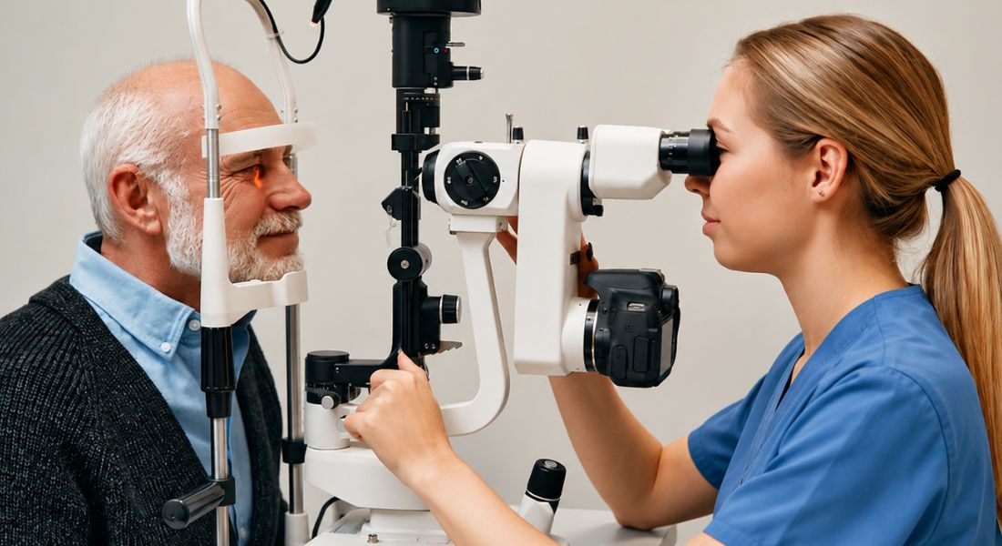
Digital slit lamp technology is becoming an increasingly important part of modern ophthalmic practice. A slit lamp is a key examination tool used by eye specialists to closely inspect the structures of the eye. Recent developments, including systems such as the Elara 900 digital slit lamp, highlight how imaging and documentation are being integrated into routine clinical assessment. These advances aim to improve the way eye examinations are recorded, shared, and reviewed over time, particularly in specialist settings.
- Enhancing Traditional Eye Examination: A slit lamp allows clinicians to examine the front of the eye in detail, including the cornea, lens, and eyelids. In glaucoma care, it also supports assessment of the drainage angle and overall eye health.
- Improved Imaging and Documentation: Digital slit lamp systems can capture high-quality images and videos during examination. This allows for more detailed documentation and easier comparison of findings over time.
- Better Communication and Teaching: Captured images can be shared with colleagues or used for teaching purposes. This supports clearer communication between clinicians and improves training opportunities for eye care professionals.
- Supporting Long-Term Monitoring: Digital records make it easier to track subtle changes in the eye over time. This can be particularly useful in chronic conditions such as glaucoma, where long-term monitoring is essential.
Digital slit lamp technology represents a meaningful step forward in how eye examinations are performed and recorded. By combining traditional clinical examination with high-quality imaging, it supports better documentation, communication, and long-term monitoring. While the core clinical skills of eye examination remain essential, these digital tools enhance accuracy and continuity of care. For patients, this means more detailed and traceable records that support ongoing eye health management.
3D Digital Surgical Visualisation
Surgical visualisation is an evolving area of innovation in ophthalmology. Haag-Streit UK has noted that the METIS 900 microscope system, featuring a new 3D Digital Heads-Up Module, is expected to be showcased at the RCOphth 2026 Congress. This reflects growing interest in advanced imaging technologies within the operating theatre.
In glaucoma surgery, high-quality visualisation is essential because procedures can be highly delicate and technically demanding. Structures such as the trabecular meshwork, drainage pathways, and surrounding tissues require precise identification and careful manipulation. Digital heads-up systems may enhance depth perception, improve ergonomics for surgeons, and allow clearer shared viewing during training and teaching.
For patients, this type of innovation is not simply about introducing new technology. It is about improving surgical precision, supporting surgeon comfort and control, and enhancing education within the operating room. Over time, these improvements can contribute to safer procedures and more consistent surgical outcomes in glaucoma care.
Smartphone-Based and Portable Imaging
Portable imaging is becoming an increasingly important area in glaucoma detection, particularly in settings where access to specialist ophthalmology services may be limited or delayed. Recent reporting has described AI-supported smartphone fundus camera systems that demonstrated high sensitivity and specificity in identifying patients who required referral for possible glaucoma in validation studies.
Although this specific development was not directly presented at the Royal College of Ophthalmologists (RCOphth) Congress, it reflects the type of innovation that is increasingly relevant to UK ophthalmology discussions. National meetings such as RCOphth frequently explore how emerging technologies can improve early detection, streamline referral pathways, and expand access to specialist assessment.
For glaucoma care, portable imaging technologies may eventually play a role in community-based screening, initial triage, and earlier identification of at-risk patients. This could help reduce delays in diagnosis and ensure that individuals who need specialist assessment are referred more efficiently into hospital-based glaucoma services.
Visual Field Testing Improvements
Visual field testing remains a fundamental part of glaucoma assessment, as it helps measure how the condition affects peripheral (side) vision. Despite its importance, the test can be tiring for patients, and results may sometimes vary between visits due to fatigue, attention, or learning effects.
Newer approaches and technologies are being developed to make visual field testing more efficient, reliable, and patient-friendly. At ophthalmology congresses such as RCOphth, discussions often focus on how to improve test accuracy and how visual field data can be better integrated with OCT imaging and clinical examination findings to provide a more complete picture of disease status.
This is particularly important because glaucoma progression is not determined by a single test result. Instead, clinicians assess patterns of change over time, combining repeated visual field tests with imaging and optic nerve evaluation to determine whether the disease is stable or worsening.
Data-Driven Glaucoma Monitoring
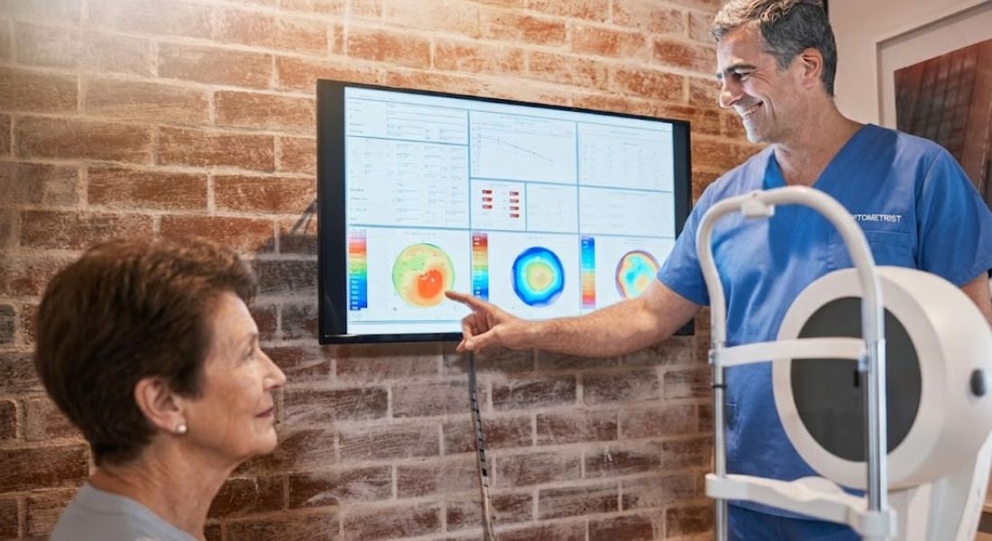
This condition generates a large amount of clinical data over time, including intraocular pressure measurements, OCT scans, visual field test results, optic nerve imaging, medication history, and any surgical interventions. Managing and interpreting this information accurately is essential for effective long-term care.
At national meetings such as the Royal College of Ophthalmologists (RCOphth) Congress, data-driven care is an increasingly relevant topic, particularly given the growing demand on UK ophthalmology services. Digital tools and integrated data systems are being explored as ways to help clinicians organise complex patient information more efficiently and identify meaningful patterns of change.
Such systems may support better prioritisation of care by highlighting patients who require urgent review due to suspected progression, while allowing those with stable disease to be monitored safely at routine intervals. This approach has the potential to improve the use of specialist time and ensure that patients receive more timely and appropriate glaucoma care.
Remote Monitoring and Virtual Clinics
Remote monitoring and virtual glaucoma clinics are becoming increasingly important within UK eye care services. For some stable patients, a full face-to-face consultation may not be required at every visit, but ongoing monitoring is still essential to ensure the disease remains stable and well controlled.
Modern digital systems can support this model by allowing clinicians to review intraocular pressure measurements, OCT imaging, visual field results, and other clinical data remotely. Consultant-led decision-making can then be carried out without every patient needing to follow the same traditional clinic pathway, helping to streamline care delivery.
However, this approach must be implemented carefully. Glaucoma is a progressive condition, and subtle signs of deterioration must not be missed. Robust systems, clear protocols, and reliable data interpretation are essential to ensure patient safety remains the highest priority.
At major ophthalmology congresses, discussions around virtual care often focus on how to balance efficiency with safety. The goal is to improve service capacity and reduce waiting times while still maintaining high-quality, consistent monitoring for all glaucoma patients.
Innovations in Eye Pressure Measurement
Eye pressure remains one of the most important modifiable risk factors in glaucoma. However, a single intraocular pressure (IOP) reading taken during a clinic visit does not always provide a complete picture of how pressure behaves throughout the day or over longer periods.
Newer approaches to pressure measurement include improved tonometry devices, discussions around home or community-based monitoring, and emerging systems designed to capture IOP fluctuations at different times. These developments aim to address the challenge that glaucoma patients may experience variations in eye pressure that are not always detected during standard clinic appointments.
Although many of these technologies are still evolving, they reflect an important clinical question: how can doctors gain a more accurate and continuous understanding of pressure control in everyday life? Better insight into pressure patterns may help improve risk assessment and treatment planning.
For patients, advances in pressure monitoring could eventually support more personalised glaucoma management, allowing treatment decisions to be better tailored to individual disease behaviour rather than relying on isolated measurements alone.
MIGS and Surgical Device Innovation
Minimally invasive glaucoma surgery (MIGS) continues to be an important area of surgical innovation in modern glaucoma care. These procedures are designed to lower intraocular pressure using techniques that generally involve less tissue disruption than traditional glaucoma operations.
At UK ophthalmology congresses, MIGS is frequently discussed in the context of patient selection, combination with cataract surgery, potential reduction in medication burden, and overall surgical safety. A key focus of these discussions is not simply whether a device or technique is new, but whether it provides meaningful and reliable benefit for appropriately selected patients in real clinical practice.
MIGS can be helpful for certain individuals, particularly those with mild to moderate glaucoma or those undergoing cataract surgery, where additional pressure lowering may be beneficial. However, it is not suitable for all patients, especially where more advanced disease requires greater pressure reduction.
Ultimately, these discussions reinforce the importance of personalised decision-making, ensuring that the chosen surgical approach aligns with disease severity, target pressure, and long-term treatment goals.
Drug Delivery Technologies
Many people with glaucoma rely on daily eye drops for long periods of time, sometimes for life. While these medications are effective, consistent use can be challenging. Difficulties may include remembering doses, applying drops correctly, or managing side effects such as irritation. Because of this, improving how medication is delivered to the eye has become an important area of research and clinical development.
- Reducing Dependence on Daily Eye Drops: New drug delivery approaches aim to lower the need for frequent drop use. This may help improve adherence, especially for patients who find daily routines difficult to maintain.
- Sustained-Release and Longer-Acting Treatments: Technologies such as sustained-release implants and longer-acting formulations are being developed. These methods aim to provide more stable drug levels over time with fewer applications.
- Improving Treatment Consistency: One of the main goals of advanced delivery systems is to make treatment more reliable. When medication is delivered in a controlled way, it reduces the risk of missed doses and fluctuating eye pressure.
- Supporting Real-World Patient Needs: For glaucoma specialists, treatment success depends not only on effectiveness but also on usability. A therapy must be practical for patients to follow in everyday life in order to work properly.
Drug delivery technologies represent an important shift in glaucoma care by focusing on improving how treatments are administered, not just what is prescribed. These developments aim to make therapy more consistent, convenient, and reliable for long-term use. While daily eye drops remain a cornerstone of treatment, newer delivery systems may help reduce treatment burden for some patients in the future. Ultimately, the goal is to ensure that glaucoma therapy is both effective and realistically manageable in everyday life.
Better Technology for Treatment Adherence
Adherence refers to how consistently patients follow their prescribed treatment. In glaucoma care, this is particularly important because missed eye drops or irregular use can lead to rises in eye pressure and an increased risk of disease progression.
Emerging technologies are being developed to support patients in maintaining regular treatment routines. These may include digital reminder systems, connected devices that track drop usage, and newer sustained-release therapies designed to reduce or replace the need for daily eye drops. Together, these innovations aim to address some of the practical challenges associated with long-term glaucoma management.
For patients, the goal of these developments is not to increase complexity or create additional pressure, but to make treatment more manageable in everyday life. By simplifying routines and supporting consistent medication use, these approaches may help improve long-term disease control and reduce the burden of ongoing glaucoma therapy.
Imaging for the Drainage Angle
The drainage angle of the eye plays a key role in glaucoma because it determines how fluid exits the eye and helps regulate intraocular pressure. Understanding the structure and function of this angle is essential for correctly diagnosing the type of glaucoma and guiding appropriate treatment decisions.
Modern imaging technologies, such as anterior segment optical coherence tomography (AS-OCT), are increasingly being used to provide detailed visualisation of the eye’s front structures. These techniques can complement traditional clinical examination methods like gonioscopy by offering additional information about angle anatomy and potential blockages.
Improved angle assessment helps specialists make more informed decisions about treatment options, including whether laser therapy, medications, minimally invasive procedures, or more traditional surgery may be most appropriate for a particular patient.
Training Technology and Simulation
The Royal College of Ophthalmologists (RCOphth) Congress plays an important role in education, training, and professional development. The 2025 Congress was described as emphasising education, collaboration, and innovation, with a strong focus on hands-on learning and simulation-based training across ophthalmology.
In glaucoma care, simulation technology can be particularly valuable because it allows surgeons to practise delicate and complex surgical steps in a controlled environment before performing them on patients. It can also support trainees in developing essential skills such as clinical examination techniques, interpretation of imaging like OCT and visual fields, and decision-making in both medical and surgical management.
Good Simulation tools help build confidence, improve technical accuracy, and standardise learning experiences. Most importantly, it supports patient safety by ensuring that clinicians are better prepared before carrying out procedures in real clinical settings, where precision and judgement are critical.
Innovation in UK Glaucoma Services
Innovation in glaucoma care is not only about new drugs, lasers, or surgical devices. It is also about how services are organised and delivered in real-world clinical settings. In the UK, glaucoma services are under increasing pressure due to rising patient numbers, long-term follow-up requirements, and the need to monitor patients with varying levels of risk. Because of this, conferences and professional discussions often focus on improving service design as much as introducing new technology.
- Improving Patient Flow and Triage Systems: Better triage systems help identify which patients need urgent review and which can be safely monitored at longer intervals. This ensures that specialist time is focused on those at highest risk of vision loss.
- Expanding Imaging and Diagnostic Pathways: Modern glaucoma services increasingly rely on imaging tools such as OCT scans and visual field testing. Streamlined pathways help ensure these tests are used efficiently and results are reviewed in a timely way.
- Development of Virtual Clinics: Virtual glaucoma clinics allow stable patients to be monitored using tests and imaging without always requiring a face-to-face appointment. This can improve capacity while maintaining safe surveillance for many patients.
- Shared Care Models Between Professionals: Collaboration between ophthalmologists, optometrists, and community eye services helps distribute workload more effectively. Shared care models can improve access and ensure patients are reviewed at appropriate intervals.
Innovation in UK glaucoma services is increasingly focused on how care is delivered, not just on new treatments. By improving triage, expanding imaging pathways, and developing virtual and shared care models, services can become more efficient and responsive. These changes are especially important in a condition like glaucoma, where long-term monitoring is essential to prevent avoidable vision loss. For patients, this means more structured, timely, and accessible care that is better matched to individual risk levels.
What These Innovations Mean for Patients
Patients do not need to understand every technical detail presented at the Royal College of Ophthalmologists (RCOphth) Congress. However, it is useful to recognise that glaucoma care is evolving across several important areas of diagnosis, monitoring, and treatment.
Clinicians are increasingly discussing advances in imaging, smarter data systems, artificial intelligence–assisted diagnosis, digital monitoring tools, surgical technologies, drug delivery systems, and remote care pathways. Although these developments vary in stage and application, they all share a common aim: improving earlier detection, enhancing the accuracy of monitoring, and supporting more personalised treatment decisions.
If you are considering glaucoma treatment in London, it is important to look for care that combines access to modern technology with careful specialist judgement. While innovation plays a valuable role, the most effective glaucoma management still depends on experienced clinical assessment, regular follow-up, and treatment plans tailored to the individual patient.
Why Clinical Judgement Still Matters
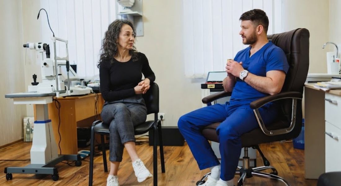
Technology plays an increasingly important role in glaucoma care, but it cannot replace a specialist’s clinical judgement. Individual test results such as scans, intraocular pressure readings, or AI-generated outputs must always be interpreted within the broader context of the patient’s overall eye health and disease history.
Factors such as age, optic nerve appearance, visual field changes, family history, corneal thickness, symptoms, treatment tolerance, and overall risk of progression all contribute to clinical decision-making. Effective glaucoma management depends on combining these elements with imaging and diagnostic technology to form a complete and accurate assessment.
This is why learning from congresses such as the RCOphth Annual Congress is so valuable. These meetings help specialists not only stay updated on new tools and technologies, but also understand how to apply them safely and appropriately in real-world clinical practice.
FAQs:
- What is the Royal College of Ophthalmologists Annual Congress?
It is a major UK ophthalmology meeting where eye specialists, researchers, and clinicians share updates on diagnosis, treatment, surgery, and innovation in eye care, including glaucoma. - Why is glaucoma a key focus at the RCOphth Congress?
Glaucoma requires lifelong monitoring and treatment, so new technologies and clinical approaches are regularly discussed to improve early detection, progression tracking, and long-term vision preservation. - What new glaucoma technologies are discussed at the Congress?
Topics often include OCT imaging advancements, artificial intelligence tools, digital slit lamps, visual field testing improvements, remote monitoring systems, and surgical innovations. - How does artificial intelligence help in glaucoma care?
AI can assist in analysing scans, detecting early structural changes, identifying progression patterns, and supporting clinicians in making more consistent and data-driven decisions. - What role does OCT imaging play in glaucoma management?
OCT provides detailed imaging of the optic nerve and retinal nerve fibre layer, helping specialists detect early damage and monitor disease progression over time. - Are new technologies replacing traditional glaucoma tests?
No. New technologies are designed to support and enhance traditional methods like eye pressure testing, visual field analysis, and clinical examination—not replace them. - What is the benefit of digital slit lamp technology?
Digital slit lamps allow high-quality imaging and recording of eye examinations, improving documentation, comparison over time, and communication between clinicians. - How is glaucoma surgery evolving at RCOphth Congress discussions?
Surgical discussions focus on minimally invasive glaucoma surgery (MIGS), improved visualisation systems, better safety outcomes, and more personalised patient selection. - What is the importance of remote monitoring in glaucoma?
Remote and virtual clinics allow stable patients to be monitored through imaging and test data without every visit needing a face-to-face appointment, improving efficiency and access. - What do these innovations mean for glaucoma patients?
They support earlier diagnosis, more accurate monitoring, improved treatment options, and more personalised care, but specialist judgement remains essential for safe management.
Final Thoughts: The Role of Innovation in Modern Glaucoma Care
The Royal College of Ophthalmologists Annual Congress highlights how rapidly glaucoma care is evolving, with innovation spanning imaging, artificial intelligence, surgical technology, drug delivery systems, and service delivery models. These advances are helping clinicians detect glaucoma earlier, monitor progression more accurately, and refine treatment decisions to better match individual patient needs. However, they are designed to complement, not replace, careful clinical assessment and long-term specialist follow-up.
Ultimately, the most important message for patients is that effective glaucoma management still relies on a combination of modern technology and experienced clinical judgement. While new tools and techniques continue to improve what is possible in diagnosis and treatment, personalised care and regular monitoring remain central to protecting vision over time. If you’re considering glaucoma treatment in London and want to know if it’s the right option, you’re welcome to reach out to us at Eye Clinic London to book a consultation.
References:
- Ang, B.C.H. et al., 2023. Recent advancements in glaucoma surgery a review of trabeculectomy, drainage devices and minimally invasive approaches. Journal of Clinical Medicine (MDPI). Available at: https://www.mdpi.com/2306-5354/10/9/1096
- Koziorowska, A.M., Opala, A. & Grabska‑Liberek, I., 2025. Non‑contact laser therapy for glaucoma: A review of direct selective laser trabeculoplasty (DSLT). Journal of Clinical Medicine. Available at: https://www.mdpi.com/2077-0383/14/19/6884
- American Academy of Ophthalmology, 2011. Laser trabeculoplasty for open‑angle glaucoma: systematic evidence and outcomes. Survey of Ophthalmology. Available at: https://www.sciencedirect.com/science/article/pii/S0161642011004611
- Gedde, S.J., 2025. Glaucoma surgery: from traditional trabeculectomy to novel minimally invasive glaucoma surgery (MIGS). Ophthalmology Clinics. Available at: https://www.sciencedirect.com/science/article/abs/pii/S2589419625001218
- Systematic review of micropulse laser trabeculoplasty (MLT) in primary open‑angle glaucoma management. Journal of Glaucoma. Available at: https://pubmed.ncbi.nlm.nih.gov/39857796/