

What Is the Difference Between Corneal Topography and Tomography?
When you start looking into laser eye surgery, you quickly realise that the consultation process involves far more than just checking your prescription. I remember when I first explored this topic, I was surprised by how many different scans and tests were needed just to properly assess suitability. It is a much more detailed process than most people expect at the beginning. Every test has a specific purpose in building a complete picture of your eye.
Two of the most important investigations you will come across are corneal topography and corneal tomography. At first, they sound very similar, and it is easy to assume they do the same thing. In reality, they provide different types of information about your cornea. Both are essential for ensuring laser eye surgery is planned safely and accurately.
Corneal topography focuses mainly on the surface of your cornea. It creates a detailed map showing the shape, curves, and contours of the outer layer. This helps your surgeon understand how the surface of your eye affects your vision. It is especially useful for identifying surface irregularities.
Corneal tomography goes deeper by analysing both the surface and the internal structure of your cornea. It creates a 3D image that shows thickness and hidden structural details that cannot be seen from the surface alone. When used together, topography and tomography give your surgeon a complete understanding of your eye, allowing for safer and more precise treatment planning.
Why Corneal Imaging Matters Before Surgery
Before any laser eye procedure, your surgeon needs a complete understanding of the shape, structure, and stability of your cornea. This is because your cornea is responsible for focusing light correctly onto the retina, and even very small irregularities can affect your vision quality. Since laser eye surgery reshapes this delicate tissue, precise planning is essential for safety and effectiveness.
Corneal imaging is therefore a crucial step in the assessment process. Without it, surgeons would not have enough detailed information about your eye’s exact structure, which would significantly increase uncertainty and risk. Imaging removes this guesswork by providing clear and measurable data before any treatment decisions are made.
Both corneal topography and tomography work together to remove that uncertainty. They allow your surgeon to understand both the surface and internal structure of your cornea in detail. This combined view helps ensure your treatment is accurately planned and tailored to your eyes.
What Is Corneal Topography?
Corneal topography focuses on the surface of your cornea. It is essentially a detailed map that shows the shape and curvature of the outer layer of your eye. This helps your surgeon understand how the surface structure may be affecting your vision. It is an important part of the pre-surgery assessment process.
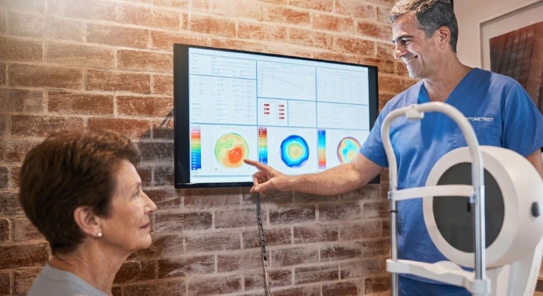
It works by projecting light patterns onto your cornea and measuring how those patterns reflect back. The collected data is then processed into a colour-coded map. This makes it easier to interpret the shape of your cornea at a glance. Even small variations in curvature can be clearly seen through this method.
Steeper areas of the cornea appear in different colours compared to flatter regions, making irregularities easy to identify. The test is quick, painless, and completely non-invasive. Despite its simplicity, it provides highly valuable information for planning laser eye surgery.
What Is Corneal Tomography?
Corneal tomography is an advanced imaging technique that provides a much deeper analysis than corneal topography. While topography focuses mainly on the surface shape of the cornea, tomography examines the entire three-dimensional structure. This includes both the front surface and the internal layers, giving a far more complete picture of corneal health. It is a key tool in modern laser eye surgery assessments because it helps detect issues that might otherwise go unnoticed.
- Full Corneal Thickness Measurement: Tomography provides highly accurate measurements of corneal thickness across the entire eye. This allows surgeons to identify variations in thickness that may affect surgical safety. It also helps in planning how much tissue can be safely removed during procedures.
- Internal Corneal Curvature Analysis: Unlike surface-only imaging, tomography evaluates the curvature of the internal corneal layers. This is important because structural changes beneath the surface can impact vision quality and surgical outcomes. It gives surgeons a deeper understanding of how the cornea behaves as a whole structure.
- Assessment of Structural Stability: Tomography helps determine whether the cornea is structurally stable over time. A stable cornea is essential for ensuring long-term success after laser eye surgery. Any signs of weakness or progressive change may influence the choice of treatment.
- Detection of Hidden Abnormalities: One of the most important benefits of tomography is its ability to reveal hidden conditions that are not visible on the surface. Early signs of diseases such as keratoconus can often be detected through this imaging. This allows surgeons to avoid unsafe procedures and recommend safer alternatives.
In conclusion, corneal tomography provides a comprehensive view of both the surface and internal structure of the eye. It offers far more detail than surface-based imaging alone, making it essential for accurate surgical planning. By revealing thickness, curvature, and hidden abnormalities, it greatly improves diagnostic precision. Ultimately, this technology plays a crucial role in ensuring laser eye surgery is both safe and appropriately tailored to each patient.
The Key Difference in Simple Terms
If I had to explain it in the simplest way, corneal topography shows the surface shape of your cornea, while corneal tomography shows the full structure from front to back. Both are used together to give your surgeon a complete understanding of your eye before any treatment is planned. They focus on different levels of detail, but both are equally important for safety and accuracy.
Topography is like looking at a landscape from above. It shows the surface curves, slopes, and irregularities of your cornea. This helps identify any uneven areas that may affect how light enters your eye. It is mainly focused on the outer surface.
Tomography, on the other hand, is like viewing a full 3D model of that same landscape, including what is beneath the surface. It shows the internal structure and thickness of your cornea as well. This deeper view helps detect hidden issues that cannot be seen with surface mapping alone.
Why You Need Both Tests
You might wonder why both corneal topography and tomography are needed if they seem to overlap. Although they are related, they provide different types of information about your cornea. Each test highlights details that the other cannot fully show. This makes them both essential for accurate laser eye surgery planning.
Topography focuses mainly on the surface of your cornea. It helps identify irregularities such as astigmatism patterns and uneven curvature on the outer layer. These surface details are important because they affect how light enters and focuses in your eye. However, it does not show what is happening beneath the surface.
Tomography looks deeper by analysing the internal structure of your cornea. It can detect issues such as thinning or hidden structural weakness that may not be visible on surface scans. When both tests are used together, they give your surgeon a complete and reliable picture of your eye health.
How Topography Works in Practice
Corneal topography is a quick and straightforward test that is commonly performed during a pre-surgery assessment. It helps your surgeon understand the exact shape of your cornea by creating a detailed surface map. The process is completely non-invasive and usually takes only a few moments to complete. It provides essential information that helps determine whether laser eye surgery is suitable for you.
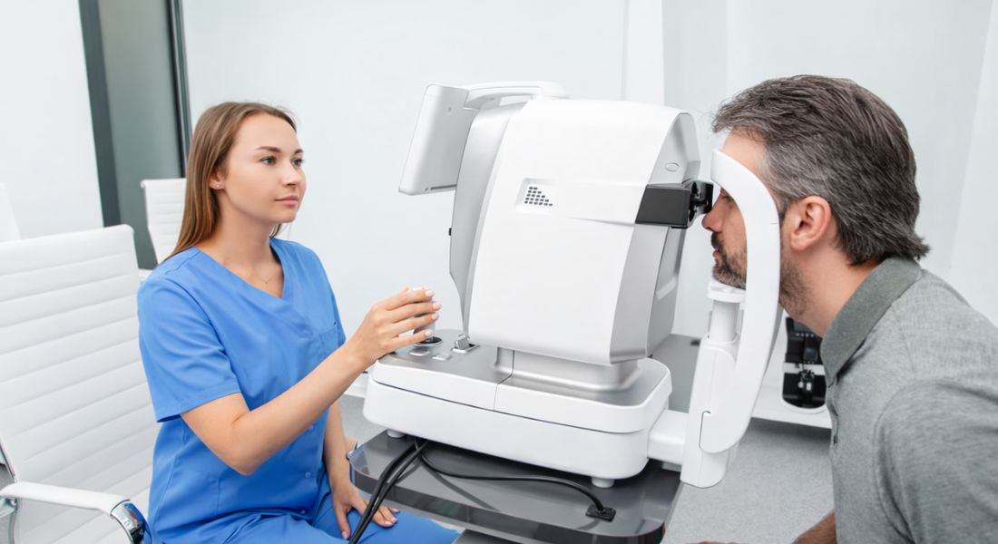
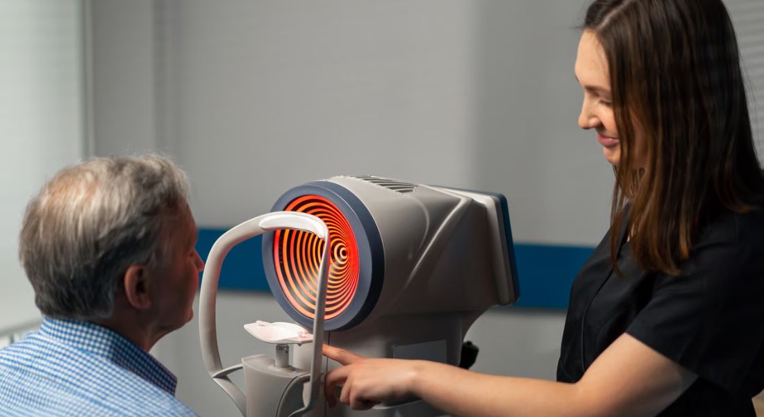
- Positioning and Fixation on a Target: You will be seated in front of a specialised scanning device and asked to focus on a small light target. This helps keep your eye steady during the imaging process. Maintaining focus ensures the machine can capture accurate and reliable data.
- Rapid Image Capture of the Cornea: The device then takes multiple images of your cornea within a few seconds. These images are captured from different angles to build a complete surface profile. The process is fast, safe, and does not cause any discomfort.
- Creation of a Detailed Corneal Map: The collected images are processed using advanced software to generate a colour-coded map of your cornea. This map shows variations in curvature across the entire surface. It allows surgeons to easily identify smooth, steep, or irregular areas.
- Assessment for Surgical Suitability: Your surgeon carefully reviews the corneal map to evaluate whether your eye is suitable for laser reshaping. They look for regularity, symmetry, and any signs of abnormal curvature. This information plays a key role in selecting the safest and most appropriate treatment option.
In conclusion, corneal topography is a fast and highly informative procedure used to assess the surface shape of the eye. It involves simple positioning, quick image capture, and advanced digital mapping. The results provide surgeons with essential insight into corneal regularity and suitability for surgery. Ultimately, this step helps ensure that any laser treatment is planned with precision and safety in mind.
How Tomography Works in Practice
Tomography uses advanced imaging technology to scan your eye in detailed layers. Instead of only looking at the surface, it captures information from different depths within the cornea. This allows it to build a complete and accurate picture of your eye’s internal structure. It is a more advanced form of assessment than surface-only scans.
It works by creating a 3D reconstruction of your cornea using the data collected during the scan. This model shows both the outer surface and the internal layers together. Your surgeon can view the cornea from multiple angles and levels of depth. This helps provide a clearer understanding of its true shape and structure.
With this information, your surgeon can measure corneal thickness across different points and detect subtle structural changes. It is especially useful for identifying early signs of conditions that may affect surgical safety. By spotting these issues early, treatment can be adjusted to ensure safer and more suitable outcomes.
Why Surface Shape Alone Is Not Enough
It might seem like the surface shape of your cornea is the most important factor, but in reality it only tells part of the story. While surface mapping is useful, it does not show what is happening beneath the outer layer. Your cornea has depth and internal structure that also need to be understood. Both levels of information are important for safe planning.
Two people can have very similar corneal surface maps but completely different internal structures. One may have a stable cornea, while the other may have early signs of thinning or weakness. These differences are not always visible from the surface alone. This is why deeper imaging is necessary before making any surgical decisions.
Without tomography, these hidden structural differences could easily be missed during assessment. This could lead to incomplete information being used when planning treatment. That is why relying on topography alone is not enough for safe laser eye surgery. Both surface and internal scans are needed to ensure a full and accurate understanding of your eye.
Detecting Early Corneal Conditions
One of the most important advantages of corneal tomography is its ability to identify early signs of corneal weakness before they become clinically obvious. Many corneal conditions develop gradually and may not immediately affect your vision in a noticeable way. This means a person can feel their eyesight is normal while subtle structural changes are already occurring beneath the surface. Early detection is therefore crucial in ensuring safe and effective treatment planning.
- Identifying Hidden Structural Changes: Tomography can detect small alterations in the cornea that are not visible during a standard eye examination. These changes may involve slight thinning or irregular internal structure. Recognising them early helps prevent progression to more serious conditions.
- Detecting Conditions Before Symptoms Appear: Some corneal disorders, such as early keratoconus, do not show clear symptoms in their initial stages. A patient may still have good vision even though the cornea is beginning to weaken. Tomography allows these early warning signs to be picked up long before noticeable vision problems develop.
- Monitoring Subtle Progression Over Time: By comparing scans over time, surgeons can observe whether the cornea is remaining stable or showing signs of change. Even small shifts in structure can be significant when planning surgery. This ongoing monitoring helps ensure that treatment decisions are based on reliable, up-to-date information.
- Guiding Safer Treatment Decisions: When early abnormalities are detected, surgeons can adjust the treatment plan accordingly. This may involve postponing surgery, choosing a different procedure, or recommending alternative vision correction options. The goal is always to prioritise long-term corneal stability and patient safety.
In conclusion, corneal tomography plays a vital role in detecting early and otherwise hidden corneal conditions. It provides detailed insights into structural changes that may not yet affect vision but are important for surgical planning. By identifying these issues early, surgeons can make more informed and safer decisions. Ultimately, this helps ensure that any treatment is tailored to protect both vision quality and long-term eye health.
How These Tests Influence Your Eligibility
Your eligibility for laser eye surgery is not based on your prescription alone. Instead, it depends heavily on the results of your corneal imaging tests. These scans give your surgeon a detailed understanding of your cornea’s shape, thickness, and overall stability. This information is essential for making a safe and accurate decision.
If your topography shows irregular surface patterns, or your tomography reveals thinning or structural instability, certain procedures may not be suitable for you. These findings help your surgeon understand whether your cornea can safely handle laser treatment. In some cases, this may limit your options or lead to a recommendation for a different approach. Every decision is based on protecting your eye health.
This is not a negative outcome, even if it feels disappointing at first. It is a safety-focused decision designed to protect your long-term vision. By identifying risks early, your surgeon can avoid procedures that may compromise your eye health in the future. This careful screening ensures that only the safest and most appropriate treatments are considered for you.
The Role of Corneal Thickness
Corneal thickness is one of the most important measurements taken during tomography. It helps your surgeon understand whether your eye can safely undergo laser reshaping. This is because the cornea must have enough structural strength to remain stable after tissue is removed. Without this safety check, planning would not be precise.
Your cornea needs to be thick enough to safely withstand the changes made during surgery. If too much tissue is removed, it can weaken the overall structure of the eye. This may affect the stability of your vision in the long term. That is why thickness is carefully evaluated before any decision is made.
Tomography plays a key role in ensuring this risk is properly assessed. It measures corneal thickness in detail and helps identify any areas that may be thinner than expected. This allows your surgeon to plan treatment more safely and accurately. It ensures that your eye remains structurally sound after the procedure.
How Surgeons Combine Both Results
Surgeons do not interpret corneal topography and tomography separately. Instead, they combine both sets of results to form a complete understanding of your eye. This ensures that every decision is based on full and accurate information. It is an essential part of safe laser eye surgery planning.
Topography provides detailed information about the surface shape of your cornea. It highlights curvature patterns and refractive issues that affect how light is focused in your eye. This helps your surgeon understand the external structure and how it relates to your vision. However, it only represents the surface layer.
Tomography adds depth by showing the internal structure and thickness of the cornea. It provides important safety information, including signs of thinning or instability that are not visible on the surface. When both tests are combined, they give a complete picture of your eye. This allows for precise and personalised surgical planning.
Why Accuracy Matters So Much
Laser eye surgery is highly precise, but that precision depends entirely on the quality of the data collected before treatment. Your surgeon relies on detailed measurements of your cornea to plan every step of the procedure. If the initial data is inaccurate, even slightly, it can affect how the treatment is designed. This is why pre-surgery testing is so important.
Even small measurement errors can influence the final outcome of surgery. A minor difference in corneal shape or thickness can change how much tissue is safely removed. This can have an impact on both safety and visual results. For this reason, every scan must be as accurate and consistent as possible.
Advanced imaging systems are used to ensure that every detail of your cornea is captured correctly. These tools detect even very small variations that are not visible in a standard eye test. Better data leads to better planning, and better planning leads to more predictable and safer visual outcomes.
What You Experience During These Tests
From your perspective, both corneal topography and tomography are very simple and comfortable procedures. You do not feel any pain or discomfort during the scans. The process is designed to be quick and easy, even if it is your first time having these tests. Most people find it far less intimidating than they expect.
The scans only take a few minutes to complete. You will be asked to look at a target light while the machine captures detailed images of your eye. There is no need for any special preparation during the scan itself. Everything is done quickly and efficiently in a clinical setting.
There is no contact with your eye at all, and nothing touches the surface of your cornea. Because of this, there is also no recovery time required afterwards. You can return to your normal activities immediately after the appointment. Most people are surprised by how straightforward and stress-free the entire process is.
How These Tests Improve Safety
Safety is the number one priority in laser eye surgery, and both corneal topography and tomography are designed with this in mind. These tests give your surgeon a detailed understanding of your cornea before any treatment is planned. By assessing both the surface and internal structure, they help ensure that no important detail is missed. This creates a much safer foundation for decision-making.
Working together, these scans help identify potential issues early, before surgery takes place. This can include surface irregularities, thinning, or signs of structural weakness that may not be visible during a basic eye exam. By detecting these factors in advance, your surgeon can plan more carefully and reduce unnecessary risk. It also helps improve the accuracy of the overall assessment.
If any concerns are found, your surgeon can avoid unsuitable procedures and recommend safer alternatives instead. In some cases, surgery may be adjusted or even delayed to protect your eye health. This is a proactive approach that prioritises long-term safety over short-term convenience. It ensures your treatment is always chosen with your best interests in mind.
How Technology Has Improved Eye Mapping
Eye imaging technology has improved dramatically over the years, making corneal assessment far more accurate and reliable. Modern devices can quickly capture detailed information about the shape and structure of your cornea. This allows surgeons to understand your eye in much greater depth than was previously possible. It has significantly improved the overall planning process for laser eye surgery.
Today’s systems provide far more detail and accuracy than older technologies. They are able to create highly detailed maps and 3D models of your cornea. These images show even small variations in curvature and structure that would have been difficult to detect before. This helps ensure that every aspect of your eye is carefully evaluated.
These advanced tools can also detect microscopic changes in the cornea that were once impossible to identify. This includes very small differences in thickness and structural integrity. As a result, potential risks can be identified earlier and taken into account during planning. This has made laser eye surgery safer and more predictable than ever before.
Choosing the Right Clinic for Assessment
Not all clinics use the same level of diagnostic technology, and this can make a significant difference to the quality of your assessment. When considering laser eye surgery, it is important to choose a clinic that invests in advanced imaging systems and carries out thorough pre-treatment testing. This ensures your corneal shape, thickness, and stability are properly understood before any decision is made. A careful approach at this stage directly supports safer outcomes.
For example, reputable providers such as Eye Clinic London place strong emphasis on detailed corneal evaluation as part of their standard assessment process. This includes advanced scans like topography and tomography, which help build a complete picture of your eye health. By using these technologies, surgeons can make more informed and accurate decisions. It also ensures that every patient is assessed individually rather than using a generalised approach.
This level of assessment ensures that your treatment is based on precise and detailed information rather than assumptions. It helps identify the most suitable procedure for your specific eye structure and reduces unnecessary risk. In turn, this leads to safer planning and more predictable results. Choosing a clinic that prioritises thorough diagnostics is therefore essential for good long-term outcomes.
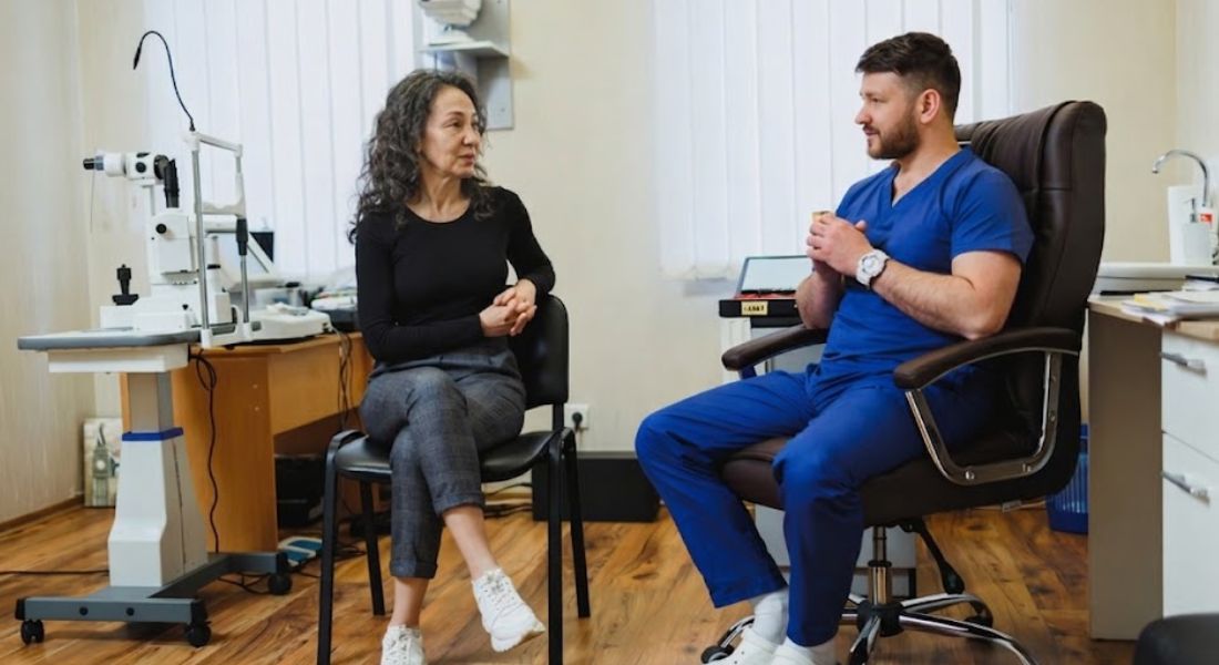
Why Understanding This Difference Helps You
Understanding the difference between topography and tomography gives you a much clearer idea of what actually happens before laser eye surgery. It shows that the process is not just a quick vision correction decision, but a detailed medical evaluation. Each test has a specific role in building a full understanding of your eye. This helps remove confusion and uncertainty from the process.
It also helps you appreciate that laser eye surgery is not only about improving vision, but also about carefully analysing and protecting your eye structure. Surgeons are not just looking at how to correct your prescription, but also at whether your cornea can safely support the procedure. This is why multiple scans are carried out before any treatment is recommended. Safety and long-term stability are always part of the decision-making process.
When you understand what is being checked and why, you are in a stronger position to make informed decisions. You can ask better questions and better understand the advice you are given. This makes the consultation process more meaningful and less overwhelming. Ultimately, it helps you feel more confident about your options and choices.
FAQs:
- What is the difference between corneal topography and tomography?
Corneal topography maps only the surface of the cornea, showing its shape, curves, and surface irregularities. Corneal tomography goes deeper by analysing both the surface and internal structure in 3D. Together, they provide a complete picture of your cornea for safer laser eye surgery planning. - Why are both topography and tomography needed before laser eye surgery?
Both tests are needed because they provide different but complementary information. Topography shows surface shape, while tomography reveals internal structure and thickness. Using both ensures your surgeon has a full understanding of your eye before making treatment decisions. - Is corneal topography enough on its own for surgery planning?
No, topography alone is not sufficient because it only shows the surface of the cornea. It cannot detect internal issues such as thinning or structural weakness. Tomography is required to complete the assessment and ensure surgical safety. - What does corneal topography show?
Corneal topography shows a colour-coded map of the cornea’s surface curvature. It highlights steep, flat, or irregular areas that may affect vision. This helps identify conditions like astigmatism and guides surface-level surgical planning. - What does corneal tomography measure?
Corneal tomography measures the full 3D structure of the cornea, including thickness and internal layers. It can detect hidden abnormalities that are not visible on the surface. This makes it essential for assessing long-term corneal stability. - Is corneal mapping painful or invasive?
No, both topography and tomography are completely painless and non-invasive. You simply look at a target while the machine scans your eye. The process takes only a few minutes and does not touch your eye. - Can tomography detect eye conditions early?
Yes, tomography can detect early signs of conditions such as keratoconus before symptoms appear. It identifies subtle structural changes that may not yet affect vision. This allows for safer surgical decisions and early intervention if needed. - How do these scans affect my eligibility for laser eye surgery?
Your eligibility depends on both surface and internal corneal health. If scans show irregularities, thinning, or instability, certain procedures may not be suitable. In such cases, safer alternatives may be recommended to protect your vision. - Why is corneal thickness important in tomography?
Corneal thickness determines whether your eye can safely undergo laser reshaping. If too much tissue is removed, it can weaken the cornea. Tomography helps measure thickness accurately to ensure enough structure remains after surgery. - What happens after corneal topography and tomography?
Your surgeon reviews both sets of results along with other tests like prescription and eye health. These findings are combined to create a personalised treatment plan. Based on the results, you may be recommended LASIK, PRK, another procedure, or no surgery if it is not safe.
Final Thoughts: Why Both Topography and Tomography Matter for Safe Laser Eye Surgery
Understanding the difference between corneal topography and tomography helps you see how detailed and carefully planned laser eye surgery really is. Topography gives your surgeon a precise map of the cornea’s surface, while tomography goes deeper to reveal the internal structure and thickness. Together, they ensure that no important detail about your eye is missed before treatment is considered.
This combined approach is what allows surgeons to design safe, personalised treatment plans. It helps identify irregularities, hidden weaknesses, and structural concerns that could affect both suitability and long-term results. Rather than relying on a single scan, your surgeon builds a complete 3D understanding of your eye to make sure the procedure is both accurate and safe. If you’re considering laser surgery in London and want to know if it’s the right option, you’re welcome to reach out to us at Eye Clinic London to book a consultation.
References:
- Kanclerz, P., Khoramnia, R. and Wang, X. (2021) Current developments in corneal topography and tomography, Diagnostics, 11(8), p. 1466. Available at: https://www.mdpi.com/2075-4418/11/8/1466
- Fan, R., Chan, T.C., Prakash, G. and Jhanji, V. (2018) Applications of corneal topography and tomography: a review, Clinical and Experimental Ophthalmology, 46(2), pp. 133–146. PubMed PMID: 29266624. Available at: https://pubmed.ncbi.nlm.nih.gov/29266624/
- Heidari, Z., Hashemi, H. & Mohammadpour, M. (2021) Evaluation of corneal topographic, tomographic and biomechanical indices for detecting clinical and subclinical keratoconus: a comprehensive three-device study. International Journal of Ophthalmology, 14(2), pp.228–239. Available at: https://pmc.ncbi.nlm.nih.gov/articles/PMC7840368/
- Klyce, S.D. (1996) Corneal topography: a review of terms and concepts. Journal of Cataract & Refractive Surgery, 22(5), pp.624–629. Available at: https://www.sciencedirect.com/science/article/abs/pii/S0886335096800228
- Razak, N., Ali, B.M. & Wan Abdul Halim, W.H. (2025) Evaluation of corneal tomographic, biomechanical and pachymetric characteristics in patients with keratoconus, their first-degree relatives, and normal individuals. Clinical Ophthalmology. Available at: https://pmc.ncbi.nlm.nih.gov/articles/PMC13034747/