

New Technologies for Keratoconus: Modern Treatment Innovations

If you’ve been living with keratoconus or you’ve just been diagnosed, you’ve probably realised that this condition affects much more than the shape of your cornea. It affects how you see, how comfortable your eyes feel and how confident you feel navigating your day-to-day life. What can feel worrying at first becomes far more manageable when you understand that keratoconus treatment has evolved dramatically. Today, you have access to technologies that didn’t exist even a few years ago technologies that improve vision, slow progression, predict disease behaviour and personalise care in ways we couldn’t imagine before.
This is an incredibly exciting time for keratoconus patients. Modern treatments are smarter, safer and more effective. Specialists can map your cornea in microscopic detail, detect early-stage disease long before symptoms appear and choose treatments tailored exactly to your eye’s unique structure. In fact, many people today avoid corneal transplants entirely thanks to new innovations.
Whether you’re exploring treatment options, considering new lenses, or simply wanting to understand what’s possible, this guide will give you a clear, comprehensive picture of the advancements transforming keratoconus care right now. My goal is to help you feel informed, reassured and empowered by the technology available to you.
Understanding Why Technology Matters in Keratoconus
Keratoconus is a condition where the structure, thickness and curvature of the cornea all change over time. That means the more precisely we can measure, diagnose, treat and stabilise the cornea, the better your long-term outcomes will be.
Modern technology helps by:
- detecting keratoconus earlier
- identifying progression sooner
- slowing or stopping weakening of the cornea
- creating smoother, clearer optical surfaces
- improving night vision and reducing glare
- offering more comfortable and precise lenses
- reducing the need for corneal transplants
- personalising treatment for each patient
This level of personalisation is one of the biggest breakthroughs in keratoconus management.
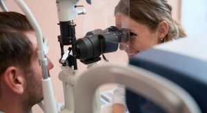
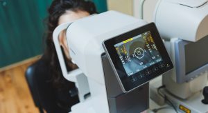
Breakthrough 1: Advanced Corneal Mapping (Topography & Tomography)
Corneal mapping technology has become one of the most powerful tools in diagnosing and managing keratoconus. Older diagnostic tools provided limited information, but today’s systems give incredibly detailed, 3D-accurate pictures of the cornea.
Placido-disc Topography
This maps the front surface of the cornea. It reveals irregularity, steepness and early shape changes.
Scheimpflug Tomography
This takes 3D cross-sectional images of the cornea. It measures both the front and back surfaces, giving a full picture of structural integrity.
OCT-Based Corneal Imaging
Optical coherence tomography allows ultra-thin slice imaging, revealing microscopic corneal details.
Why this matters:
These technologies detect keratoconus years earlier than older tools. Early detection allows early cross-linking, which stops or slows progression.
Breakthrough 2: Epithelial Thickness Mapping
Epithelial mapping is one of the newest, most powerful diagnostic innovations.
The epithelium the thin outer layer of the cornea changes thickness depending on what is happening beneath it. When keratoconus forms a cone, the epithelium becomes thinner over the cone and thicker around it.
This pattern is highly sensitive and reveals:
- very early keratoconus
- progression before vision changes
- the exact location of corneal weakness
- suitability for different procedures
Why it’s important:
This has revolutionised early diagnosis, especially for children and teens who progress faster.
Breakthrough 3: Corneal Cross-Linking (CXL) New Protocols & Devices
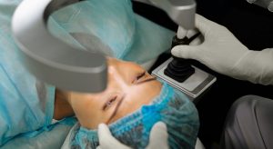
Cross-linking remains one of the most important treatment breakthroughs in keratoconus. But recent innovations have made CXL faster, safer and more customisable.
Today’s improvements include:
Accelerated CXL – Uses stronger UV light in shorter sessions, reducing treatment time.
Customised CXL – Targets only the weakened areas of the cornea, sparing healthy tissue.
Transepithelial (Epi-On) CXL –Performs CXL without removing the epithelium, reducing discomfort and recovery time.
Pulsed UV Light Protocols – Helps oxygen regenerate more effectively for better results.
Why this matters:
Cross-linking is now more comfortable, faster and available to more patients, including those who previously weren’t good candidates.
Breakthrough 4: Topography-Guided Laser Treatments
Laser treatments for keratoconus are not about thinning the cornea they’re about smoothing the surface to improve clarity. Topography-guided procedures have become revolutionary for selected patients.
Examples include:
- topography-guided PRK
- Contoura Vision
- customised ablation patterns
These treatments smooth irregularities to:
- reduce ghosting
- improve night vision
- sharpen clarity
- improve lens tolerance
- reduce glare and starbursts
Often, these are combined with cross-linking for stability.
Why this matters:
Many patients previously considered unsuitable for laser treatments can now benefit thanks to customised topography-guided techniques.
Breakthrough 5: Intracorneal Ring Segments (ICRS) Smarter Designs & Techniques
Ring segments have evolved significantly.
Modern rings come in:
- different thicknesses
- different curvatures
- precise arc lengths
- improved materials
Used with femtosecond lasers for exact placement, these implants help:
- flatten steep corneas
- improve symmetry
- reduce irregular astigmatism
- enhance contact lens comfort
- delay or avoid transplants
Why this matters:
Newer ring designs offer better outcomes and more predictable results.
Breakthrough 6: Modern Scleral Lenses A Game Changer
Scleral lenses have become one of the most important innovations for keratoconus patients. They vault over the cornea and rest on the sclera, creating a smooth optical surface.
Today’s scleral lens technologies include:
High-oxygen materials
Allow long, comfortable wear.
Customised digital lens mapping
Matches your exact corneal shape using 3D modelling.
Wavefront-guided optics
Reduces subtle distortions and halos.
Tear reservoir control
Improves hydration and comfort throughout the day.
Expanded diameter options
Optimised for extremely irregular corneas.
Why this matters:
Scleral lenses restore clarity for many moderate to advanced keratoconus patients who struggle with soft lenses or glasses.
Breakthrough 7: Hybrid Contact Lenses
Hybrid lenses combine the best of both worlds:
- a rigid centre for clarity
- a soft skirt for comfort
These lenses are great for patients who:
- cannot tolerate rigid lenses
- need more clarity than soft lenses
- want more stability during sports
- need a balance between comfort and performance
Modern hybrid designs have improved tear exchange and fit.
Breakthrough 8: Custom Soft Keratoconus Lenses
New soft lens designs are made specifically for keratoconus.
These lenses:
- have thicker centres for stability
- use advanced optics to reduce halos
- offer improved comfort for sensitive eyes
- help mild to moderate keratoconus patients
These are excellent for people who want the familiarity of soft lenses with the clarity of rigid optics.
Breakthrough 9: Artificial Intelligence (AI) in Keratoconus Diagnosis
AI is changing everything about how keratoconus is detected and monitored.
AI algorithms analyse:
- shape patterns
- thickness maps
- epithelial distribution
- progression trends
- risk profiles
This helps predict:
- which patients will progress
- how quickly progression will occur
- which treatments will work best
- early signs before clinical changes appear
Why this matters:
AI-guided diagnosis catches crises before they happen.
Breakthrough 10: Genetic & Molecular Research
Scientists are discovering genetic markers associated with keratoconus.
This research aims to:
- predict risk earlier
- identify at-risk family members
- create targeted drug therapies
- understand how inflammation contributes to weakening
While still in development, these innovations will reshape treatment in the future.
Breakthrough 11: Bowman Layer Transplantation
This new technique involves transplanting the Bowman layer a thin corneal layer into the weak cornea.
Benefits include:
- strengthening the cornea
- reducing steepness
- improving long-term stability
- delaying full transplants
It’s ideal for patients with advanced keratoconus who still want to avoid a corneal graft.
Breakthrough 12: Modern Corneal Transplants (PK & DALK Improvements)
Transplants are now safer, stronger and more durable.
Improvements include:
- femtosecond laser-assisted graft shaping
- zig-zag incision patterns for stability
- improved suturing techniques
- stronger donor tissue processing
- faster recovery times
These refinements reduce rejection risk and improve vision quality.
Breakthrough 13: Smart Monitoring Tools for Home Use
New digital tools allow you to track your vision from home.
Apps and devices can track:
- corneal curvature
- visual distortion patterns
- light scatter
- night vision patterns
- daily fluctuations
These tools help determine exactly when clinical intervention is needed.
Breakthrough 14: Combining Technologies for Personalised Care
The biggest innovation in keratoconus isn’t one technology it’s the combination of many.
Today specialists can mix:
- cross-linking
- topography-guided laser
- intracorneal rings
- scleral lenses
- hybrid lenses
- customised soft lenses
This creates a fully personalised plan that treats not just the cornea but also your lifestyle needs.
This approach was impossible a decade ago.
How These Innovations Improve Real-Life Outcomes
The technologies available today offer life-changing improvements:
- clearer day and night vision
- reduced ghosting
- better driving confidence
- smoother reading experiences
- fewer headaches from eye strain
- longer comfortable lens wear
- fewer transplants
- stronger, more stable corneas
- earlier detection for children and teens
- increased independence
Keratoconus no longer means surrendering your quality of life. You now have options.
Daily Life with Modern Keratoconus Technology
Modern keratoconus treatments and technologies are not just about improving clinical measurements they genuinely transform how you move through your day. From driving to working on screens, and even enjoying hobbies, today’s advanced lenses and personalised treatments make daily tasks far more comfortable and predictable.
Driving: Scleral lenses customised soft lenses, and topography-guided treatments can significantly reduce glare, distortion, and ghosting on the road. Many patients notice more stable vision during bright daylight, and improved clarity when reading road signs or navigating traffic at night.
Working: Custom lens designs created using corneal maps offer sharper, more stable vision for screen-heavy work. This reduces the strain that comes from blurry text or fluctuating focus, making long hours at a computer much more manageable.
Sports: Modern scleral lenses and hybrid lenses provide a secure, stable fit that stays in place even during fast movement. Whether you enjoy running, cycling, or indoor sports, these designs help maintain crisp vision without the irritation or shifting common with older lenses.
Reading: Improved optical quality from wavefront-guided lenses and advanced cross-linking techniques helps reduce ghosting, double images, and shadowing around letters. This makes reading books, documents, and even phone screens far more comfortable.
Night-Time Vision: Wavefront-guided optics and refined lens technology help control halos, starbursts, and streaks around lights. These improvements make night driving, evening walks, or using digital screens in dim lighting much easier and safer.
Frequently Asked Questions:
- Can keratoconus be completely cured with new technology?
No, keratoconus cannot be “cured” in the traditional sense, but modern technology can effectively stop progression, improve vision and prevent severe complications. Treatments like corneal cross-linking strengthen the cornea to halt worsening, while topography-guided laser procedures and advanced lenses significantly improve clarity. With today’s innovations, many patients maintain excellent long-term vision without ever needing a corneal transplant. - Is corneal cross-linking still necessary with all these new innovations?
Cross-linking remains the only treatment proven to stop keratoconus from progressing, so it is still a cornerstone of care even with newer technologies. What has changed is how cross-linking is performed. Newer accelerated, pulsed, customised and transepithelial methods make the procedure faster, safer and more comfortable while offering the same or even better strengthening outcomes. - Are topography-guided laser treatments safe for keratoconus patients?
Yes, they are safe for carefully selected patients when performed by a specialist experienced in keratoconus management. These treatments do not try to reshape the cornea like standard LASIK. Instead, they gently smooth irregularities on the surface to improve clarity, reduce glare and enhance lens tolerance. They are often paired with cross-linking for added stability. - Are scleral lenses difficult to wear or maintain?
Most patients adjust quickly to scleral lenses because they rest on the white part of the eye, not the sensitive cornea. They often feel more comfortable than rigid lenses and provide superior clarity. Insertion and removal require a short learning period, but once mastered, most people find them easy to manage. Modern high-oxygen materials also make them safe for long daily wear. - How early can keratoconus be detected with new imaging tools?
Advanced mapping tools like tomography, epithelial thickness mapping and AI-based analysis can detect keratoconus years before symptoms appear. These technologies identify subtle structural changes that older tools missed. Early detection is crucial because it allows doctors to perform cross-linking while the cornea is still strong, preventing major progression. - Will I still need a corneal transplant with all these new treatment options?
Most patients no longer require a corneal transplant thanks to cross-linking, intracorneal ring segments, advanced lenses and modern laser options. Even those with more advanced disease may benefit from Bowman layer transplantation as a less invasive alternative. Transplants are now a last resort rather than a common step in treatment. - What role does artificial intelligence play in keratoconus management?
AI analyses thousands of data points from corneal scans to predict which patients are at highest risk of progression and how rapidly the disease may change. It helps clinicians choose the best treatment before problems arise. AI also improves diagnostic accuracy, reduces misdiagnosis and ensures that early keratoconus cases especially in children are not overlooked. - Can children and teenagers benefit from these new technologies?
Yes, and in fact, early intervention is even more important for younger patients because their keratoconus tends to progress faster. Technologies like epithelial thickness mapping, tomography and AI tools make early detection possible, while accelerated and transepithelial cross-linking provide safer, quicker treatment options with minimal discomfort. These innovations significantly reduce the need for future transplants. - Are newer contact lens designs better than traditional rigid lenses?
Absolutely. While rigid gas-permeable lenses were once the standard, today’s options scleral lenses, hybrid lenses and customised soft keratoconus lenses offer far better comfort, clarity and stability. Modern lens technology is designed to match the unique shape of each cornea using 3D data, which means fewer fittings, a more natural visual experience and reduced irritation or dryness. - What is the future of keratoconus treatment?
The future is moving toward fully personalised care combining genetic testing, AI-driven predictions, targeted cross-linking, custom lenses and early disease detection. Researchers are developing molecular treatments that may one day strengthen the cornea without surgery. Imaging technology is becoming even more precise, allowing specialists to monitor tiny structural changes over time. As these innovations evolve, keratoconus will become even more manageable, with an increasing number of patients living normal, glasses-free lives.
Final Thoughts: Your Next Step Toward Clearer Vision
Keratoconus care has progressed further in the last decade than in the previous fifty years. With advanced imaging, personalised cross-linking, modern lens technologies, and AI-supported diagnostics, patients today have more control, better vision, and far more treatment options than ever before. If you’re exploring your next steps and want expert guidance tailored to your eyes, it’s always best to speak directly with a specialist who understands the full spectrum of these innovations. If you’re considering Keratoconus treatment in London and want to know whether it’s the right choice for your situation, you’re welcome to reach out to us at Eye Clinic London to book a consultation.
References:
- Santodomingo-Rubido, J., Huseynova, T., Soria, F., Hartwig, A. & Limbek, H. (2022) ‘Keratoconus: an updated review’, Contact Lens & Anterior Eye. https://www.sciencedirect.com/science/article/pii/S1367048421002058
- Bui, A. D. et al. (2023) ‘Keratoconus Diagnosis and Treatment: Recent Advances and Emerging Needs’, Clinical Ophthalmology, [online] https://pmc.ncbi.nlm.nih.gov/articles/PMC10511017/
- Morales, P., et al. (2025) ‘Advances in Intracorneal Ring Segment (ICRS) implantation for keratoconus’, Journal of Clinical Medicine, 14(13), 4454. https://www.mdpi.com/2077-0383/14/13/4454
- Rafizadeh, S. M., et al. (2025) ‘Keratoconus and quality of life: an updated comprehensive review’, Clinical Ophthalmology, [online] https://pmc.ncbi.nlm.nih.gov/articles/PMC12395790/
- The Evolution of Diagnostics for Keratoconus Bevara, A. et al. (2023). This review describes advances in corneal tomography and topography that have improved accuracy and early detection of keratoconus. https://pubmed.ncbi.nlm.nih.gov/36598277/